Explore
 Validate
 Learn
Learn Western blot
 Immunohistochemistry
 Chromatin Immunoprecipitation
Antibody data
- Antibody Data
- Antigen structure
- References [1]
- Comments [0]
- Validations
- Immunohistochemistry [1]
- Flow cytometry [10]
- Other assay [1]
Submit
Validation data
Reference
Comment
Report error
- Product number
- MA5-16171 - Provider product page

- Provider
- Invitrogen Antibodies
- Product name
- DNMT3A Monoclonal Antibody (64B1446)
- Antibody type
- Monoclonal
- Antigen
- Recombinant full-length protein
- Description
- In Western blot, this antibody detects a band at approximately 120 kDa (predicted molecular weight: 102 kDa). Staining of formalin-fixed tissues is enhanced by boiling tissue sections in 10 mM sodium citrate buffer, pH 6.0 for 10-20 min followed by cooling at RT for 20 min. Suggested positive control: antigen standard for DNMT3A (transient overexpression lysate), transfected cell lysate (WB), HeLa cells (ICC).
- Reactivity
- Human, Mouse, Rat
- Host
- Mouse
- Isotype
- IgG
- Antibody clone number
- 64B1446
- Vial size
- 100 μg
- Concentration
- 1 mg/mL
- Storage
- Store at 4°C short term. For long term storage, store at -20°C, avoiding freeze/thaw cycles.
Submitted references DNMT1-Mediated DNA Methylation Targets CDKN2B to Promote the Repair of Retinal Ganglion Cells in Streptozotocin-Induced Mongolian Gerbils during Diabetic Retinopathy.
Wang X, Zhang J, Liao Y, Jin Y, Yu X, Li H, Yang Q, Li X, Chen R, Wu D, Zhu H
Computational and mathematical methods in medicine 2022;2022:9212116
Computational and mathematical methods in medicine 2022;2022:9212116
No comments: Submit comment
Supportive validation
- Submitted by
- Invitrogen Antibodies (provider)
- Main image

- Experimental details
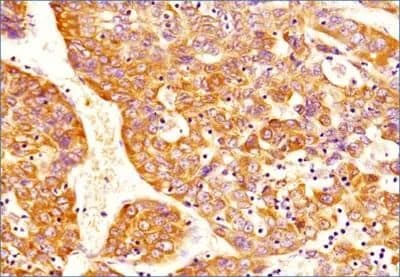
- Immunohistochemical analysis of DNMT3A in human hepatocellular carcinoma tissue section. Samples were incubated in DNMT3A monoclonal antibody (Product # MA5-16171) using a dilution of 1:100. The antibody generated a specific cytoplasmic staining in all the cancer cells while some of the cells depicted nuclear staining also.
Supportive validation
- Submitted by
- Invitrogen Antibodies (provider)
- Main image

- Experimental details
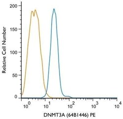
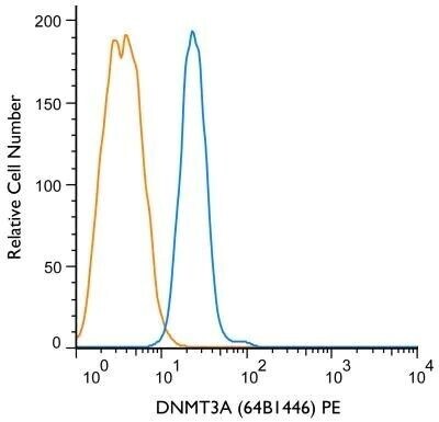
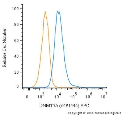
- Flow cytometry of DNMT3A in NTERA-2 cells (blue) and a matched isotype control (orange). Samples were incubated in DNMT3A monoclonal antibody (Product # MA5-16171) using a dilution of 1 µg/mL for 30 minutes at room temperature. Cells were fixed with 4% PFA and then permeablized with 0.1% saponin. Both antibodies were conjugated to Phycoerythrin.

- Submitted by
- Invitrogen Antibodies (provider)
- Main image

- Experimental details
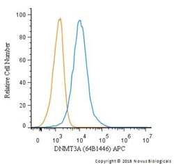
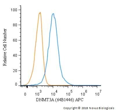
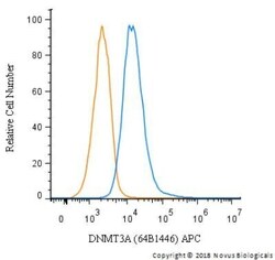
- Flow cytometry of DNMT3A in HepG2 cells. Samples were incubated in DNMT3A monoclonal antibody (Product # MA5-16171) using a dilution of 1 µg/mL for 30 minutes at room temperature. Antibody (blue) and a matched isotype control (orange). Cells were fixed with 4% PFA and then permeablized with 0.1% saponin. Both antibodies were conjugated to allophycocyanin (APC).

- Submitted by
- Invitrogen Antibodies (provider)
- Main image

- Experimental details
- Flow cytometry of DNMT3A in HeLa cells. Samples were incubated in DNMT3A monoclonal antibody (Product # MA5-16171) using a dilution of 1 µg/mL for 30 minutes at room temperature. Antibody (blue) and a matched isotype control (orange). Cells were fixed with 4% PFA and then permeabilized with 0.1% saponin. Both antibodies were conjugated to allophycocyanin (APC).

- Submitted by
- Invitrogen Antibodies (provider)
- Main image
- Experimental details
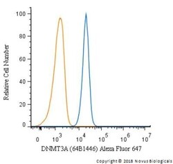
- Flow cytometry of DNMT3A in Jurkat cells. Samples were incubated in DNMT3A monoclonal (Product # MA5-16171) using a dilution of 2.5 µg/mL for 30 minutes at room temperature. Antibody (blue) and a matched isotype control (orange). Cells were fixed with 4% PFA and then permeablized with 0.1% saponin. Both antibodies were conjugated to Alexa Fluor 647.

- Submitted by
- Invitrogen Antibodies (provider)
- Main image

- Experimental details
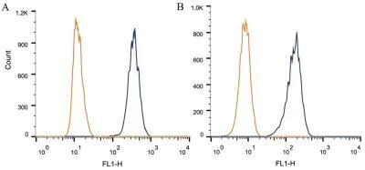
- Flow cytometry of DNMT3A in 1 x 10^6 CHO (A) and HEK-293 (B) cells. Samples were incubated in DNMT3A monoclonal antibody (Product # MA5-16171) using a dilution of 1 µg/1x10^6 cells. Antibody (dark blue). Isotype control shown in orange.

- Submitted by
- Invitrogen Antibodies (provider)
- Main image

- Experimental details
- Flow cytometry of DNMT3A in Jurkat cells. Samples were incubated in DNMT3A monoclonal (Product # MA5-16171) using a dilution of 2.5 µg/mL for 30 minutes at room temperature. Antibody (blue) and a matched isotype control (orange). Cells were fixed with 4% PFA and then permeablized with 0.1% saponin. Both antibodies were conjugated to Alexa Fluor 647.

- Submitted by
- Invitrogen Antibodies (provider)
- Main image

- Experimental details
- Flow cytometry of DNMT3A in 1 x 10^6 CHO (A) and HEK-293 (B) cells. Samples were incubated in DNMT3A monoclonal antibody (Product # MA5-16171) using a dilution of 1 µg/1x10^6 cells. Antibody (dark blue). Isotype control shown in orange.
- Submitted by
- Invitrogen Antibodies (provider)
- Main image
- Experimental details
- Flow cytometry of DNMT3A in NTERA-2 cells (blue) and a matched isotype control (orange). Samples were incubated in DNMT3A monoclonal antibody (Product # MA5-16171) using a dilution of 1 µg/mL for 30 minutes at room temperature. Cells were fixed with 4% PFA and then permeablized with 0.1% saponin. Both antibodies were conjugated to Phycoerythrin.
- Submitted by
- Invitrogen Antibodies (provider)
- Main image
- Experimental details
- Flow cytometry of DNMT3A in HepG2 cells. Samples were incubated in DNMT3A monoclonal antibody (Product # MA5-16171) using a dilution of 1 µg/mL for 30 minutes at room temperature. Antibody (blue) and a matched isotype control (orange). Cells were fixed with 4% PFA and then permeablized with 0.1% saponin. Both antibodies were conjugated to allophycocyanin (APC).
- Submitted by
- Invitrogen Antibodies (provider)
- Main image
- Experimental details
- Flow cytometry of DNMT3A in HeLa cells. Samples were incubated in DNMT3A monoclonal antibody (Product # MA5-16171) using a dilution of 1 µg/mL for 30 minutes at room temperature. Antibody (blue) and a matched isotype control (orange). Cells were fixed with 4% PFA and then permeabilized with 0.1% saponin. Both antibodies were conjugated to allophycocyanin (APC).
Supportive validation
- Submitted by
- Invitrogen Antibodies (provider)
- Main image
- Experimental details
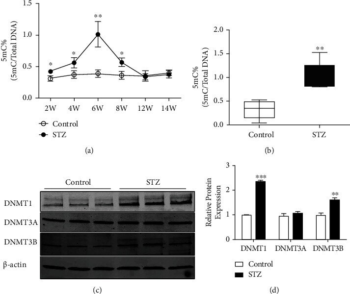
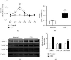
- Figure 3 DNMT1-mediated DNA methylation was significantly increased in the early phase of diabetic retinopathy. (a) STZ injection upregulated the global DNA methylation level of the retina until 6 weeks after injection. (b) Quantification of the average 5-mC content in DNA methylation of the retina isolated from sodium citrate buffer injection (control) and STZ injection (STZ) groups. (c-d) Western blot showed the protein expression levels of DNMT1, DNMT3A, and DNMT3B. * P < 0.05, ** P < 0.01.