 Explore
 Validate
 Learn
Learn Western blot
Antibody data
- Antibody Data
- Antigen structure
- References [0]
- Comments [0]
- Validations
- Western blot [4]
- Immunocytochemistry [1]
- Flow cytometry [1]
Submit
Validation data
Reference
Comment
Report error
- Product number
- MA1-41013 - Provider product page

- Provider
- Invitrogen Antibodies
- Product name
- DNMT3A Monoclonal Antibody (64B814.1)
- Antibody type
- Monoclonal
- Antigen
- Recombinant full-length protein
- Description
- Suggested positive control: antigen standard for DNMT3A (transient overexpression lysate), transfected cell lysate, mouse ES cells.
- Reactivity
- Human, Mouse
- Host
- Mouse
- Isotype
- IgG
- Antibody clone number
- 64B814.1
- Vial size
- 100 µg
- Concentration
- 1 mg/mL
- Storage
- Store at 4°C short term. For long term storage, store at -20°C, avoiding freeze/thaw cycles.
No comments: Submit comment
Supportive validation
- Submitted by
- Invitrogen Antibodies (provider)
- Main image

- Experimental details
- Western blot analysis of Dnmt3a using a DNA Methyltransferase 3a monoclonal antibody (Product # MA1-41013) at 2 µg/mL in 293 cell lysate transfected with either mouse Dnmt3a (lane 1) or mouse Dnmt3b (lane 2).

- Submitted by
- Invitrogen Antibodies (provider)
- Main image

- Experimental details
- Western blot analysis of Dnmt3a in (A) Dnmt3a transfected and (B) untransfected 293 cell lysate using a DNA Methyltransferase 3a monoclonal antibody (Product # MA1-41013) at 1 µg/mL.

- Submitted by
- Invitrogen Antibodies (provider)
- Main image

- Experimental details
- Western blot analysis of DNMT3A in 293 cell lysate. Samples were incubated in DNMT3A monoclonal antibody (Product # MA1-41013) using a dilution of 1 µg/mL.

- Submitted by
- Invitrogen Antibodies (provider)
- Main image

- Experimental details
- Western blot was performed using Anti-DNMT3A Monoclonal Antibody (64B814.1) (Product # MA1-41013) and 102 kDa, 77 kDa bands corresponding to DNMT3A isoforms along with uncharacterized bands (*) at ~50 kDa and ~18 kDa were observed in the cell lines, with higher expression in NTERA-2, NCCIT, iPSC compared to HeLa, MCF7, and Jurkat. Modified whole cell extracts (1% SDS) (40 µg lysate) of HeLa (Lane 1), MCF7 (Lane 2), Jurkat (Lane 3), NTERA-2 (Lane 4), NCCIT (Lane 5) and iPSC (Lane 6) were electrophoresed using Novex® NuPAGE® 4-12 % Bis-Tris gel (Product # NP0321BOX). Resolved proteins were then transferred onto a nitrocellulose membrane (Product # IB23001) by iBlot® 2 Dry Blotting System (Product # IB21001). The blot was probed with the primary antibody (1 µg/mL concentration) and detected by chemiluminescence with Goat anti-Mouse IgG (H+L), Superclonal™ Recombinant Secondary Antibody, HRP conjugate (Product # A28177, 1:4000 dilution) using the iBright FL 1000 (Product # A32752). Chemiluminescent detection was performed using Novex® ECL Chemiluminescent Substrate Reagent Kit (Product # WP20005).

Supportive validation
- Submitted by
- Invitrogen Antibodies (provider)
- Main image
- Experimental details
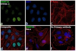
- Immunofluorescence analysis of DNMT3A was performed using 70% confluent log phase NTERA-2 cells. The cells were fixed with 4% paraformaldehyde for 10 minutes, permeabilized with 0.1% Triton™ X-100 for 15 minutes, and blocked with 2% BSA for 1 hour at room temperature. The cells were labeled with DNMT3A Monoclonal Antibody (64B814.1) (Product # MA1-41013) at 5 µg/mL in 0.1% BSA, incubated at 4 degree Celsius overnight and then with Donkey anti-Mouse IgG (H+L) Highly Cross-Adsorbed Secondary Antibody, Alexa Fluor Plus 488 (Product # A32766) at a dilution of 1:2000 for 45 minutes at room temperature (Panel a: green). Nuclei (Panel b: blue) were stained with SlowFade® Gold Antifade Mountant with DAPI (Product # S36938). F-actin (Panel c: red) was stained with Rhodamine Phalloidin (Product # R415, 1:300). Panel d represents the merged image showing nuclear localization. Panel e represents HeLa cells having no expression of DNMT3A. Panel f represents control cells with no primary antibody to assess background. The images were captured at 60X magnification.

Supportive validation
- Submitted by
- Invitrogen Antibodies (provider)
- Main image

- Experimental details
- Flow cytometry of DNMT3A in HeLa cells. Samples were incubated in DNMT3A monoclonal antibody (Product # MA1-41013). Antibody (blue) and a matched isotype control (orange). Both antibodies were conjugated to Alexa Fluor (R) 488.
