Explore
 Validate
 Learn
Learn Western blot
 Immunocytochemistry
Antibody data
- Antibody Data
- Antigen structure
- References [1]
- Comments [0]
- Validations
- Immunocytochemistry [2]
- Other assay [1]
Submit
Validation data
Reference
Comment
Report error
- Product number
- PA5-110384 - Provider product page

- Provider
- Invitrogen Antibodies
- Product name
- UCP4 Polyclonal Antibody
- Antibody type
- Polyclonal
- Antigen
- Recombinant full-length protein
- Description
- When using two or more Super Bright dye-conjugated antibodies in a staining panel, it is recommended to use Super Bright Complete Staining Buffer (Product # SB-4401) to minimize any non-specific polymer interactions. Please refer to the datasheet for Super Bright Staining Buffer for more information.
- Reactivity
- Human, Mouse, Rat
- Host
- Rabbit
- Isotype
- IgG
- Vial size
- 100 µL
- Concentration
- 1.75 mg/mL
- Storage
- -20° C, Avoid Freeze/Thaw Cycles
Submitted references In vivo magnetic resonance spectroscopy in the brain of Cdkl5 null mice reveals a metabolic profile indicative of mitochondrial dysfunctions.
Carli S, Chaabane L, Butti C, De Palma C, Aimar P, Salio C, Vignoli A, Giustetto M, Landsberger N, Frasca A
Journal of neurochemistry 2021 May;157(4):1253-1269
Journal of neurochemistry 2021 May;157(4):1253-1269
No comments: Submit comment
Supportive validation
- Submitted by
- Invitrogen Antibodies (provider)
- Main image
- Experimental details
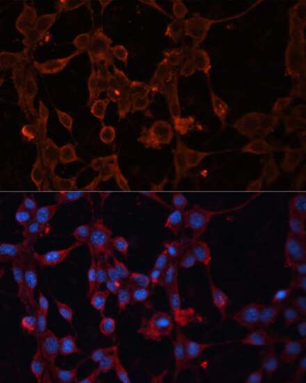
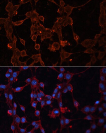
- Immunocytochemistry analysis of UCP4 in NIH/3T3 cells. Samples were incubated in polyclonal UCP4 antibody (Product # PA5-110384) using a dilution of 1:100, followed by DAPI.
- Submitted by
- Invitrogen Antibodies (provider)
- Main image
- Experimental details
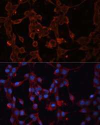
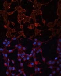
- Immunofluorescence analysis of UCP4 in NIH/3T3 cells. Samples were incubated with UCP4 Polyclonal antibody (Product # PA5-110384) using a dilution of 1:100. Blue: DAPI for nuclear staining.
Supportive validation
- Submitted by
- Invitrogen Antibodies (provider)
- Main image
- Experimental details
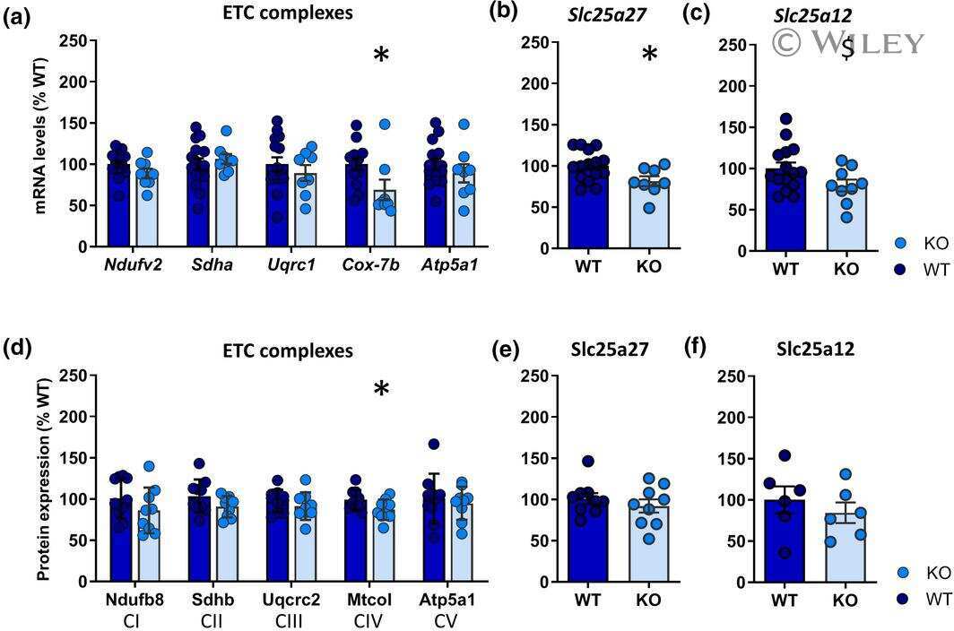
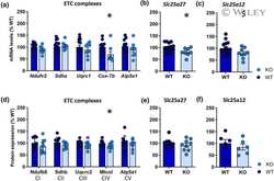
- 6 FIGURE Cdkl5 KO brains display an aberrant expression of complex IV of the electron transport chain. (a-c) The transcriptional expression of mitochondrial genes was analyzed by quantitative RT-PCR in WT and KO hippocampi ( n = 15 WT and 8 KO). Results displayed a consistent reduction of Cox7b and Slc25a27 , while Slc25a12 showed a decreasing trend. Hprt was used as internal standard. The fold changes of transcript levels, compared to WT animals, are reported as mean +- SEM ( $ p < .068; * p < .05). (d-f) Western blots of mitochondrial proteins from WT and KO mice. A significant decrease was confirmed for the complex IV of the ETC, Cox7b. Bar graphs show the mean +- SEM of the expression levels in Cdkl5 KO compared to WT, of ETCs (total OXPHOS) ( n = 10 WT, n = 9 KO), Slc25a27 (UCP4) ( n = 9 WT, n = 9 KO), and Slc25a12 (Aralar) ( n = 6 WT, n = 6 KO). Full blots are reported in Figure S1. Statistical significance was assessed by Student's t test ( Ndufv2, Sdha, Uqrc1, Atp5a1 , Slc25a27, and Slc25a12 in quantitative RT-PCR and ETC complexes and Slc25a12 in Western Blot ) or Mann-Whitney U test ( Cox7b in quantitative RT-PCR and Slc25a27 in Western Blot), in accordance with D'Agostino-Pearson test for data distribution; * p < .05