 Explore
 Validate
 Learn
Learn Western blot
Antibody data
- Antibody Data
- Antigen structure
- References [0]
- Comments [0]
- Validations
- Western blot [1]
- Immunocytochemistry [2]
- Immunohistochemistry [5]
- Flow cytometry [1]
- Protein array [1]
- Other assay [1]
Submit
Validation data
Reference
Comment
Report error
- Product number
- V7301 - Provider product page

- Provider
- NSJ Bioreagents
- Product name
- RPSA Antibody / 40S Ribosomal protein SA / Laminin Receptor 1
- Antibody type
- Monoclonal
- Description
- This highly specific RPSA antibody is suitable for use in Flow cytometry/Immunofluorescence/Western blot/Immunohistochemistry applications with human samples.
- Reactivity
- Human
- Host
- Mouse
- Conjugate
- Unconjugated
- Antibody clone number
- RPSA/2699
- Vial size
- 20 ug (with BSA and sodium azide), 100 ug (with BSA and sodium azide), 100 ug (without BSA or sodium azide), 7 ml IHC only format (if applicable)
- Concentration
- 0.2 mg/ml, 1 mg/ml
- Storage
- Store the RPSA antibody at 2-8oC (with azide) or aliquot and store at -20oC or colder (without azide).
No comments: Submit comment
Supportive validation
- Submitted by
- NSJ Bioreagents (provider)
- Main image

- Experimental details
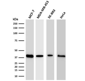
- Western blot testing of human MCF-7, MDA-MB-453, SK-BR3, and HeLa cell lysate with RPSA antibody. Routinely observed molecular weight: 37-40 kDa and 67 kDa.
Supportive validation
- Submitted by
- NSJ Bioreagents (provider)
- Main image
- Experimental details
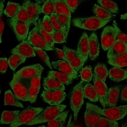
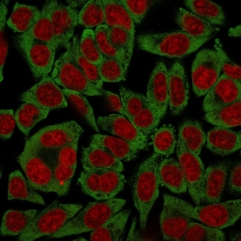
- Immunofluorescent staining of human HeLa cells with RPSA antibody (green, clone RPSA/2699) and Reddot nuclear stain (red).
- Submitted by
- NSJ Bioreagents (provider)
- Main image

- Experimental details
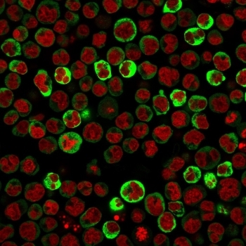
- Immunofluorescent staining of paraformaldehyde-Raji cells with RPSA antibody (green, clone RPSA/2699) and Reddot nuclear stain (red).
Supportive validation
- Submitted by
- NSJ Bioreagents (provider)
- Main image

- Experimental details
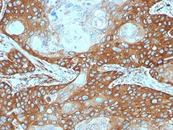
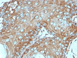
- IHC testing of FFPE human cervical carcinoma stained with RPSA antibody. Required HIER: boiling tissue sections in 10mM citrate buffer, pH6, for 10-20 min followed by cooling at RT for 20 min.
- Submitted by
- NSJ Bioreagents (provider)
- Main image
- Experimental details
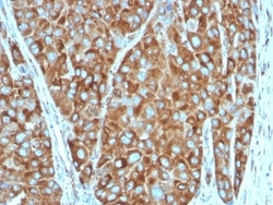
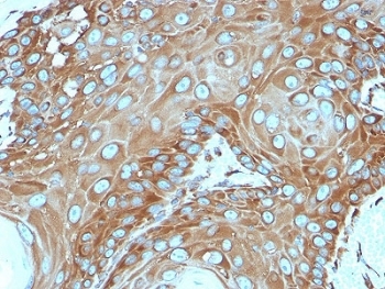
- IHC testing of FFPE human colon carcinoma stained with RPSA antibody. Required HIER: boiling tissue sections in 10mM citrate buffer, pH6, for 10-20 min followed by cooling at RT for 20 min.
- Submitted by
- NSJ Bioreagents (provider)
- Main image

- Experimental details
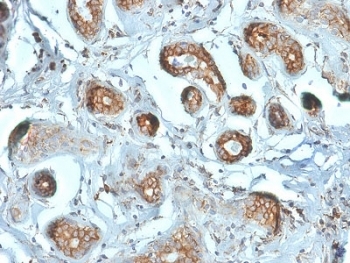
- IHC testing of FFPE human breast carcinoma stained with RPSA antibody. Required HIER: boiling tissue sections in 10mM citrate buffer, pH6, for 10-20 min followed by cooling at RT for 20 min.
- Submitted by
- NSJ Bioreagents (provider)
- Main image
- Experimental details
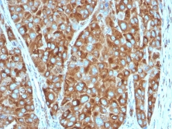
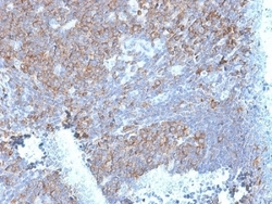
- IHC testing of FFPE human basal cell carcinoma stained with RPSA antibody. Required HIER: boiling tissue sections in 10mM citrate buffer, pH6, for 10-20 min followed by cooling at RT for 20 min.
- Submitted by
- NSJ Bioreagents (provider)
- Main image
- Experimental details
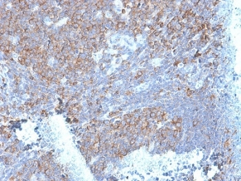
- IHC testing of FFPE human tonsil stained with RPSA antibody. Required HIER: boiling tissue sections in 10mM citrate buffer, pH6, for 10-20 min followed by cooling at RT for 20 min.
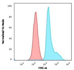
Supportive validation
- Submitted by
- NSJ Bioreagents (provider)
- Main image

- Experimental details
- FACS staining of paraformaldehyde-Raji cells with RPSA antibody (clone RPSA/2699); Red=isotype control, Blue= MSH6 antibody.
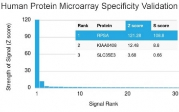
Supportive validation
- Submitted by
- NSJ Bioreagents (provider)
- Main image

- Experimental details
- Analysis of HuProt(TM) microarray containing more than 19,000 full-length human proteins using RPSA antibody. These results demonstrate the foremost specificity of the RPSA/2699 mAb.Z- and S- score: The Z-score represents the strength of a signal that an antibody (in combination with a fluorescently-tagged anti-IgG secondary Ab) produces when binding to a particular protein on the HuProt(TM) array. Z-scores are described in units of standard deviations (SD's) above the mean value of all signals generated on that array. If the targets on the HuProt(TM) are arranged in descending order of the Z-score, the S-score is the difference (also in units of SD's) between the Z-scores. The S-score therefore represents the relative target specificity of an Ab to its intended target.
Supportive validation
- Submitted by
- NSJ Bioreagents (provider)
- Main image
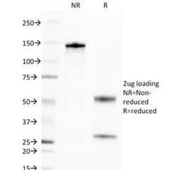
- Experimental details
- SDS-PAGE analysis of purified, BSA-free RPSA antibody as confirmation of integrity and purity.
