Explore
 Validate
 Learn
Learn Western blot
Antibody data
- Antibody Data
- Antigen structure
- References [2]
- Comments [0]
- Validations
- Western blot [6]
- Immunohistochemistry [2]
Submit
Validation data
Reference
Comment
Report error
- Product number
- NBP1-33002 - Provider product page

- Provider
- Novus Biologicals
- Proper citation
- Novus Cat#NBP1-33002, RRID:AB_2182396
- Product name
- Rabbit Polyclonal RPSA Antibody
- Antibody type
- Polyclonal
- Description
- Immunogen affinity purified.
- Reactivity
- Human, Mouse
- Host
- Rabbit
- Isotype
- IgG
- Vial size
- 0.1 ml
- Storage
- Aliquot and store at -20C or -80C. Avoid freeze-thaw cycles.
Submitted references Green Tea Extract Rich in Epigallocatechin-3-Gallate Prevents Fatty Liver by AMPK Activation via LKB1 in Mice Fed a High-Fat Diet.
Expression of 67-kDa laminin receptor was associated with tumor progression and poor prognosis in epithelial ovarian cancer.
Santamarina AB, Oliveira JL, Silva FP, Carnier J, Mennitti LV, Santana AA, de Souza GH, Ribeiro EB, Oller do Nascimento CM, Lira FS, Oyama LM
PloS one 2015;10(11):e0141227
PloS one 2015;10(11):e0141227
Expression of 67-kDa laminin receptor was associated with tumor progression and poor prognosis in epithelial ovarian cancer.
Song T, Choi CH, Cho YJ, Sung CO, Song SY, Kim TJ, Bae DS, Lee JW, Kim BG
Gynecologic oncology 2012 May;125(2):427-32
Gynecologic oncology 2012 May;125(2):427-32
No comments: Submit comment
Supportive validation
- Submitted by
- Novus Biologicals (provider)
- Main image
- Experimental details
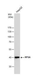
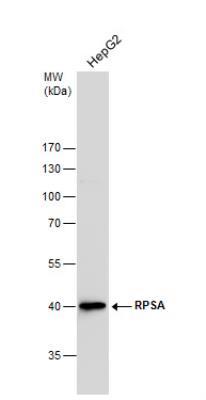
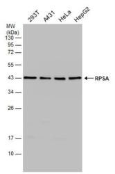
- Western Blot: RPSA Antibody [NBP1-33002] - Whole cell extracts (30 ug) was separated by 10 % SDS-PAGE, and blotted with RPSA antibody diluted by 1:1000
- Submitted by
- Novus Biologicals (provider)
- Main image

- Experimental details
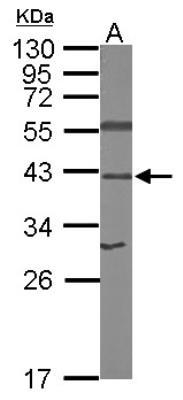
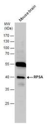
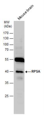
- Western Blot: RPSA Antibody [NBP1-33002] - Sample (50 ug of whole cell lysate) A: mouse brain 12% SDS PAGE; antibody diluted at 1:1000.
- Submitted by
- Novus Biologicals (provider)
- Main image
- Experimental details
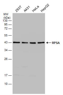
- Western Blot: RPSA Antibody [NBP1-33002] - Various whole cell extracts (30 ug) were separated by 12% SDS-PAGE, and the membrane was blotted with RPSA antibody diluted at 1:1000.
- Submitted by
- Novus Biologicals (provider)
- Main image

- Experimental details
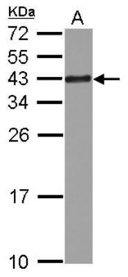
- Western Blot: RPSA Antibody [NBP1-33002] - Sample (30 ug of whole cell lysate). A: Jurkat. 12% SDS PAGE. RPSA Antibody diluted at 1:1000
- Submitted by
- Novus Biologicals (provider)
- Main image
- Experimental details
- Western Blot: RPSA Antibody [NBP1-33002] - RPSA antibody detects RPSA protein by Western blot analysis. Mouse tissue extracts (50 ug) was separated by 10 % SDS-PAGE, and the membrane was blotted with RPSA antibody diluted by 1:1000.
- Submitted by
- Novus Biologicals (provider)
- Main image
- Experimental details
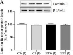
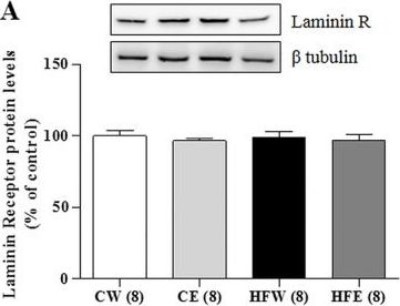
- Western Blot: RPSA Antibody [NBP1-33002] - Liver protein expression in different experimental groups of Laminin R, AdipoR2 and SIRT1.Western Blotting analysis of protein expression in the liver on different experimental groups of aminin Receptor. Image shows demonstrative bands of the analyzed proteins and respective housekeeping protein (beta-tubulin) in liver. Data are expressed in mean +/- s.e.m. *p
Supportive validation
- Submitted by
- Novus Biologicals (provider)
- Main image
- Experimental details
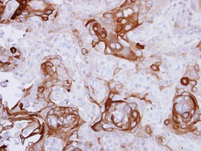
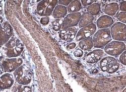
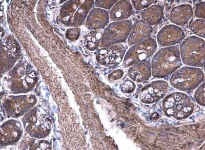
- Immunohistochemistry-Paraffin: RPSA Antibody [NBP1-33002] - Mouse small intestine. RPSA antibody [N1C3] diluted at 1:500. Antigen Retrieval: Trilogy™ (EDTA based, pH 8.0) buffer, 15min.
- Submitted by
- Novus Biologicals (provider)
- Main image
- Experimental details
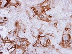
- Immunohistochemistry-Paraffin: RPSA Antibody [NBP1-33002] - CA922 xenograft, using RPSA antibody at 1:500 dilution. Antigen Retrieval: Trilogy™ (EDTA based, pH 8.0) buffer, 15min.