Explore
 Validate
 Learn
Learn Western blot
 Immunocytochemistry
Antibody data
- Antibody Data
- Antigen structure
- References [1]
- Comments [0]
- Validations
- Immunocytochemistry [2]
- Immunohistochemistry [3]
Submit
Validation data
Reference
Comment
Report error
- Product number
- MA5-26346 - Provider product page

- Provider
- Invitrogen Antibodies
- Product name
- GCLC Monoclonal Antibody (OTI1A3)
- Antibody type
- Monoclonal
- Antigen
- Recombinant full-length protein
- Reactivity
- Human, Mouse, Rat
- Host
- Mouse
- Isotype
- IgG
- Antibody clone number
- OTI1A3
- Vial size
- 100 μL
- Concentration
- 1 mg/mL
- Storage
- -20°C, Avoid Freeze/Thaw Cycles
Submitted references Spatial locations of certain enzymes and transporters within preinvasive ductal epithelial cells predict human breast cancer recurrences.
Kraft AM, Petty HR
American journal of physiology. Cell physiology 2020 Nov 1;319(5):C910-C921
American journal of physiology. Cell physiology 2020 Nov 1;319(5):C910-C921
No comments: Submit comment
Supportive validation
- Submitted by
- Invitrogen Antibodies (provider)
- Main image

- Experimental details
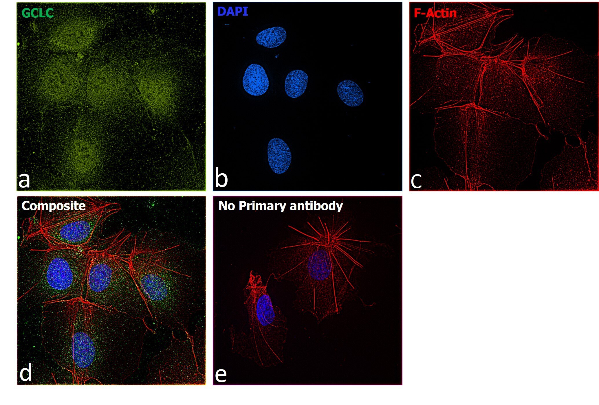
- Immunofluorescence analysis GCLC Monoclonal Antibody (OTI1A3) was performed using 70% confluent log phase HepG2 cells. The cells were fixed with 4% paraformaldehyde for 10 minutes, permeabilized with 0.1% Triton™ X-100 for 15 minutes, and blocked with 2% BSA for 1 hour at room temperature. The cells were labeled GCLC monoclonal antibody (Product # MA5-26346) at 1:100 dilution in 0.1% BSA, incubated at 4 degree Celsius overnight and then with Goat anti-Mouse IgG (H+L), Superclonal™ Recombinant Secondary Antibody, Alexa Fluor 488 (Product # A28175) at a dilution of 1:2000 for 45 minutes at room temperature (Panel a: green). Nuclei (Panel b: blue) were stained with SlowFade® Gold Antifade Mountant with DAPI (Product # S36938). F-actin (Panel c: red) was stained with Rhodamine Phalloidin (Product # R415, 1:300). Panel d represents the merged image showing nuclear localization. Panel e represents control cells with no primary antibody to assess background. The images were captured at 60X magnification.

- Submitted by
- Invitrogen Antibodies (provider)
- Main image
- Experimental details
- Immunofluorescence analysis GCLC Monoclonal Antibody (OTI1A3) was performed using 70% confluent log phase HepG2 cells. The cells were fixed with 4% paraformaldehyde for 10 minutes, permeabilized with 0.1% Triton™ X-100 for 15 minutes, and blocked with 2% BSA for 1 hour at room temperature. The cells were labeled GCLC monoclonal antibody (Product # MA5-26346) at 1:100 dilution in 0.1% BSA, incubated at 4 degree Celsius overnight and then with Goat anti-Mouse IgG (H+L), Superclonal™ Recombinant Secondary Antibody, Alexa Fluor 488 (Product # A28175) at a dilution of 1:2000 for 45 minutes at room temperature (Panel a: green). Nuclei (Panel b: blue) were stained with SlowFade® Gold Antifade Mountant with DAPI (Product # S36938). F-actin (Panel c: red) was stained with Rhodamine Phalloidin (Product # R415, 1:300). Panel d represents the merged image showing nuclear localization. Panel e represents control cells with no primary antibody to assess background. The images were captured at 60X magnification.
Supportive validation
- Submitted by
- Invitrogen Antibodies (provider)
- Main image
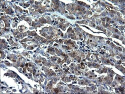
- Experimental details
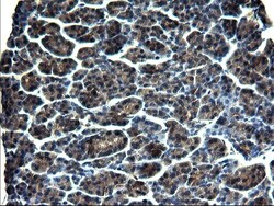
- Immunohistochemistry was performed on paraffin-embedded human pancreas tissue. To expose target proteins, 1 mM EDTA in 10mM Tris, pH8.5, 120°C for 3min was used. Following antigen retrieval, tissues were probed with a GCLC monoclonal antibody (Product # MA5-26346).

- Submitted by
- Invitrogen Antibodies (provider)
- Main image
- Experimental details
- Immunohistochemistry was performed on paraffin-embedded carcinoma of human thyroid tissue. To expose target proteins, 1 mM EDTA in 10mM Tris, pH8.5, 120°C for 3min was used. Following antigen retrieval, tissues were probed with a GCLC monoclonal antibody (Product # MA5-26346).

- Submitted by
- Invitrogen Antibodies (provider)
- Main image

- Experimental details
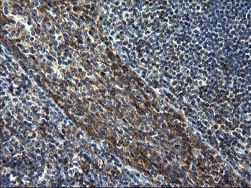
- Immunohistochemistry was performed on paraffin-embedded human lymph node tissue. To expose target proteins, 1 mM EDTA in 10mM Tris, pH8.5, 120°C for 3min was used. Following antigen retrieval, tissues were probed with a GCLC monoclonal antibody (Product # MA5-26346).