 Explore
 Validate
 Learn
Learn Western blot
Antibody data
- Antibody Data
- Antigen structure
- References [3]
- Comments [0]
- Validations
- Western blot [3]
- Immunocytochemistry [1]
Submit
Validation data
Reference
Comment
Report error
- Product number
- PA5-19702 - Provider product page

- Provider
- Invitrogen Antibodies
- Product name
- GCLC Polyclonal Antibody
- Antibody type
- Polyclonal
- Antigen
- Synthetic peptide
- Description
- This antibody is predicted to react with cow based on sequence homology.
- Reactivity
- Human, Mouse, Rat
- Host
- Rabbit
- Isotype
- IgG
- Vial size
- 100 µg
- Concentration
- 0.9 mg/mL
- Storage
- Store at 4°C short term. For long term storage, store at -20°C, avoiding freeze/thaw cycles.
Submitted references Histone Methyltransferase G9a Drives Chemotherapy Resistance by Regulating the Glutamate-Cysteine Ligase Catalytic Subunit in Head and Neck Squamous Cell Carcinoma.
Cigarette Smoke-Induced Hypermethylation of the GCLC Gene Is Associated With COPD.
Nuclear Factor, Erythroid 2-Like 2 Regulates Expression of Type 3 Inositol 1,4,5-Trisphosphate Receptor and Calcium Signaling in Cholangiocytes.
Liu CW, Hua KT, Li KC, Kao HF, Hong RL, Ko JY, Hsiao M, Kuo ML, Tan CT
Molecular cancer therapeutics 2017 Jul;16(7):1421-1434
Molecular cancer therapeutics 2017 Jul;16(7):1421-1434
Cigarette Smoke-Induced Hypermethylation of the GCLC Gene Is Associated With COPD.
Cheng L, Liu J, Li B, Liu S, Li X, Tu H
Chest 2016 Feb;149(2):474-482
Chest 2016 Feb;149(2):474-482
Nuclear Factor, Erythroid 2-Like 2 Regulates Expression of Type 3 Inositol 1,4,5-Trisphosphate Receptor and Calcium Signaling in Cholangiocytes.
Weerachayaphorn J, Amaya MJ, Spirli C, Chansela P, Mitchell-Richards KA, Ananthanarayanan M, Nathanson MH
Gastroenterology 2015 Jul;149(1):211-222.e10
Gastroenterology 2015 Jul;149(1):211-222.e10
No comments: Submit comment
Supportive validation
- Submitted by
- Invitrogen Antibodies (provider)
- Main image

- Experimental details
- Western blot analysis of Human Skeletal Muscle Tissue Lysate using Product # PA5-19702, GCLC primary antibody at a dilution of 1 µg/mL (lane 1). Staining of Human Kidney Tissue Lysate at a dilution of 1 µg/mL (lane 2). Blot treated with a secondary HRP-conjugated Goat polyclonal anti-Rabbit antibody was used at a dilution of 1:3000.

- Submitted by
- Invitrogen Antibodies (provider)
- Main image
- Experimental details
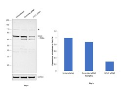
- Knockdown of GCLC was achieved by transfecting Hep G2 with GCLC specific siRNAs (Silencer® select Product # s5800). Western blot analysis (Fig. a) was performed using whole cell extracts from the GCLC knockdown cells (lane 3), non-specific scrambled siRNA transfected cells (lane 2) and untransfected cells (lane 1). The blot was probed with GCLC Polyclonal Antibody (Product # PA5-19702, 1ug/ml) and Goat anti-Rabbit IgG (H+L), Superclonal™ Recombinant Secondary Antibody, HRP (Product # A27036, 0.25µg/ml, 1:4000 dilution). Densitometric analysis of this western blot is shown in histogram (Fig. b). Decrease in signal upon siRNA mediated knock down confirms that antibody is specific to GCLC. An uncharacterized band (*) at ~110 kDa was observed in the samples.

- Submitted by
- Invitrogen Antibodies (provider)
- Main image

- Experimental details
- Western blot was performed using Anti-GCLC Polyclonal Antibody (Product # PA5-19702) and a 72 kDa band corresponding to GCLC was observed across cell lines tested along with an uncharacterized band at ~110 kDa. Whole cell extracts (30 µg lysate) of A549 (Lane 1), Hep G2 (Lane 2), MCF7 (Lane 3), Jurkat (Lane 4), Caco-2 (Lane 5), Mouse Liver (Lane 6), Mouse Heart (Lane 7) and Mouse Kidney (Lane 8) were electrophoresed using NuPAGE™ 4-12% Bis-Tris Protein Gel (Product # NP0322BOX). Resolved proteins were then transferred onto a nitrocellulose membrane (Product # IB23001) by iBlot® 2 Dry Blotting System (Product # IB21001). The blot was probed with the primary antibody (1ug/ml) and detected by chemiluminescence with Goat anti-Rabbit IgG (H+L) Superclonal™ Recombinant Secondary Antibody, HRP (Product # A27036, 1:4000 dilution) using the iBright FL 1000 (Product # A32752). Chemiluminescent detection was performed using Novex® ECL Chemiluminescent Substrate Reagent Kit (Product # WP20005).

Supportive validation
- Submitted by
- Invitrogen Antibodies (provider)
- Main image

- Experimental details
- Immunofluorescence analysis GCLC Polyclonal Antibody was performed using 70% confluent log phase A549 cells. The cells were fixed with 4% paraformaldehyde for 10 minutes, permeabilized with 0.1% Triton™ X-100 for 15 minutes, and blocked with 2% BSA for 1 hour at room temperature. The cells were labeled with GCLC Polyclonal Antibody (Product # PA5-19702) at 5 µg/mL in 0.1% BSA, incubated at 4 degree Celsius overnight and then with Goat anti-Rabbit IgG (H+L) Superclonal™ Recombinant Secondary Antibody, Alexa Fluor® 488 conjugate (Product # A27034) at a dilution of 1:2000 for 45 minutes at room temperature (Panel a: green). Nuclei (Panel b: blue) were stained with SlowFade® Gold Antifade Mountant with DAPI (Product # S36938). F-actin (Panel c: red) was stained with Rhodamine Phalloidin (Product # R415, 1:300). Panel d represents the merged image showing cytosolic localization. Panel e represents control cells with no primary antibody to assess background. The images were captured at 60X magnification.
