 Explore
 Validate
 Learn
Learn Western blot
 ELISA
ELISAAntibody data
- Antibody Data
- Antigen structure
- References [1]
- Comments [0]
- Validations
- Western blot [3]
- Immunocytochemistry [2]
- Immunohistochemistry [1]
Submit
Validation data
Reference
Comment
Report error
- Product number
- MA5-15740 - Provider product page

- Provider
- Invitrogen Antibodies
- Product name
- MSH2 Monoclonal Antibody (1B3=3A2B8C)
- Antibody type
- Monoclonal
- Antigen
- Purifed from natural sources
- Description
- MA5-15740 targets MSH2 in indirect ELISA, IF, IHC, and WB applications and shows reactivity with Human and Non-human primate samples. The MA5-15740 immunogen is purified recombinant fragment of human MSH2 expressed in E. Coli. MA5-15740 detects MSH2 which has a predicted molecular weight of approximately 105kDa.
- Reactivity
- Human
- Host
- Mouse
- Isotype
- IgG
- Antibody clone number
- 1B3=3A2B8C
- Vial size
- 100 µL
- Concentration
- Conc. Not Determined
- Storage
- Store at 4°C short term. For long term storage, store at -20°C, avoiding freeze/thaw cycles.
Submitted references Focal amplifications are associated with chromothripsis events and diverse prognoses in gastric cardia adenocarcinoma.
Zhao XK, Xing P, Song X, Zhao M, Zhao L, Dang Y, Lei LL, Xu RH, Han WL, Wang PP, Yang MM, Hu JF, Zhong K, Zhou FY, Han XN, Meng CL, Ji JJ, Chen X, Wang LD
Nature communications 2021 Nov 11;12(1):6489
Nature communications 2021 Nov 11;12(1):6489
No comments: Submit comment
Supportive validation
- Submitted by
- Invitrogen Antibodies (provider)
- Main image
- Experimental details
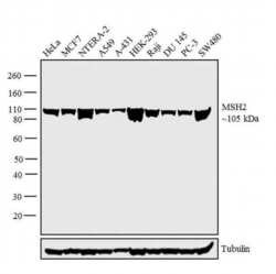
- Western blot analysis was performed on nuclear enriched extracts (30 µg lysate) of HeLa (Lane 1), MCF7 (Lane 2), NTERA-2 (Lane 3), A549 (Lane 4), A-431 (Lane 5), HEK-293 (Lane 6), Raji (Lane 7), DU 145 (Lane 8), PC-3 (Lane 9) and SW480 (Lane 10). The blot was probed with Anti-MSH2 Monoclonal Antibody (Product # MA5-15740, 1:1000 dilution) and detected by chemiluminescence using Goat anti-Mouse IgG (H+L) Superclonal™ Secondary Antibody, HRP conjugate (Product # A28177, 0.25 µg/mL, 1:4000 dilution). A 105 kDa band corresponding to MSH2 was observed across the cell lines tested.

- Submitted by
- Invitrogen Antibodies (provider)
- Main image

- Experimental details
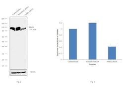
- Western blot analysis of MSH2 (Fig. a) was performed by loading 30 ug of HeLa Control (lane 1), HeLa Cas9 (lane 2) and HeLa MSH2 knockout (lane 3) modified whole cell extracts (1% SDS). MSH2 was detected at 95kDa using MSH2 Monoclonal Antibody (1B3=3A2B8C) (Product # MA5-15740, 1:1000 dilution) and Goat anti-Mouse IgG (H+L) Superclonal™ Secondary Antibody, HRP conjugate (Product # A28177, 0.25 µg/ml, 1:4000 dilution) Densitometric analysis of this western blot is shown in histogram (Fig. b). Loss of signal in CRISPR mediated knockout (KO) confirms that antibody is specific to MSH2.

- Submitted by
- Invitrogen Antibodies (provider)
- Main image
- Experimental details
- Knockdown of MSH2 was achieved by transfecting HeLa with MSH2 specific siRNAs (Silencer® select Product # s16285, s16286). Western blot analysis (Fig. a) was performed using modified whole cell extracts (1% SDS) from the MSH2 knockdown cells (lane 3), non-specific scrambled siRNA transfected cells (lane 2) and untransfected cells (lane 1). The blots were probed with MSH2 Monoclonal Antibody (1B3=3A2B8C) (Product # MA5-15740, 1:1000 dilution) and Goat anti-Mouse IgG (H+L) Superclonal™ Secondary Antibody, HRP conjugate (Product # A28177, 0.25 µg/mL, 1:4000 dilution). Densitometric analysis of this western blot is shown in histogram (Fig. b). Reduction in signal upon siRNA mediated knock down confirms that antibody is specific to MSH2.

Supportive validation
- Submitted by
- Invitrogen Antibodies (provider)
- Main image

- Experimental details
- Immunofluorescence analysis of HeLa cells using MSH2 monoclonal antibody (Product # MA5-15740) (Green). Red: actin filaments have been labeled with phalloidin.

- Submitted by
- Invitrogen Antibodies (provider)
- Main image

- Experimental details
- Immunofluorescence analysis of MSH2 was performed using 70% confluent log phase HeLa cells. The cells were fixed with 4% paraformaldehyde for 10 minutes, permeabilized with 0.1% Triton™ X-100 for 15 minutes, and blocked with 1% BSA for 1 hour at room temperature. The cells were labeled with MSH2 Mouse Monoclonal Antibody (Product # MA5-15740) at 1:100 dilution in 0.1% BSA, incubated at 4 degree Celsius overnight and then labeled with Goat anti-Mouse IgG (H+L) Superclonal™ Secondary Antibody, Alexa Fluor® 488 conjugate (Product # A28175) at a dilution of 1:2000 for 45 minutes at room temperature (Panel a: green). Nuclei (Panel b: blue) were stained with SlowFade® Gold Antifade Mountant with DAPI (Product # S36938). F-actin (Panel c: red) was stained with Rhodamine Phalloidin (Product # R415, 1:300). Panel d represents the merged image showing nuclear localization. Panel e represents control cells with no primary antibody to assess background. The images were captured at 60X magnification.

Supportive validation
- Submitted by
- Invitrogen Antibodies (provider)
- Main image

- Experimental details
- Immunohistochemical analysis of paraffin-embedded human breast cancer (left) and lung cancer (right) tissues using MSH2 monoclonal antibody (Product # MA5-15740) followed with DAB staining.
