Explore
 Validate
 Learn
Learn Western blot
 Other assay
Antibody data
- Antibody Data
- Antigen structure
- References [3]
- Comments [0]
- Validations
- Other assay [2]
Submit
Validation data
Reference
Comment
Report error
- Product number
- PA5-15177 - Provider product page

- Provider
- Invitrogen Antibodies
- Product name
- MYLK Polyclonal Antibody
- Antibody type
- Polyclonal
- Antigen
- Synthetic peptide
- Description
- This antibody is predicted to react with rabbit based on sequence homology.
- Reactivity
- Human, Mouse
- Host
- Rabbit
- Isotype
- IgG
- Vial size
- 400 μL
- Concentration
- 2 mg/mL
- Storage
- Store at 4°C short term. For long term storage, store at -20°C, avoiding freeze/thaw cycles.
Submitted references Behind Brain Metastases Formation: Cellular and Molecular Alterations and Blood-Brain Barrier Disruption.
Picturing Breast Cancer Brain Metastasis Development to Unravel Molecular Players and Cellular Crosstalk.
Listeria Adhesion Protein Induces Intestinal Epithelial Barrier Dysfunction for Bacterial Translocation.
Godinho-Pereira J, Garcia AR, Figueira I, Malhó R, Brito MA
International journal of molecular sciences 2021 Jun 30;22(13)
International journal of molecular sciences 2021 Jun 30;22(13)
Picturing Breast Cancer Brain Metastasis Development to Unravel Molecular Players and Cellular Crosstalk.
Figueira I, Galego S, Custódio-Santos T, Vicente R, Molnár K, Haskó J, Malhó R, Videira M, Wilhelm I, Krizbai I, Brito MA
Cancers 2021 Feb 22;13(4)
Cancers 2021 Feb 22;13(4)
Listeria Adhesion Protein Induces Intestinal Epithelial Barrier Dysfunction for Bacterial Translocation.
Drolia R, Tenguria S, Durkes AC, Turner JR, Bhunia AK
Cell host & microbe 2018 Apr 11;23(4):470-484.e7
Cell host & microbe 2018 Apr 11;23(4):470-484.e7
No comments: Submit comment
Supportive validation
- Submitted by
- Invitrogen Antibodies (provider)
- Main image
- Experimental details
- NULL
- Submitted by
- Invitrogen Antibodies (provider)
- Main image
- Experimental details
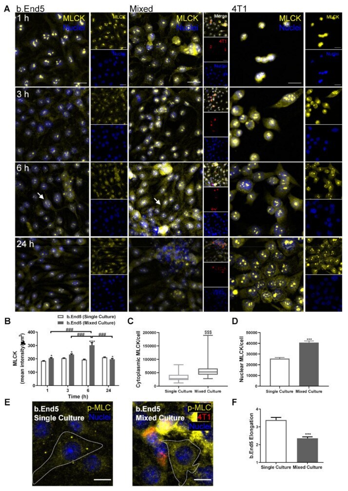
- Figure 6 Interaction between breast cancer cells and brain microvascular endothelial cells leads to cytoskeleton rearrangement. Confluent monolayers of the brain endothelioma cell line b.End5 under physiological laminar non-pulsatile shear stress were exposed to 4T1 breast cancer cells (previously labelled with CellTracker(tm) DMTPX Red Dye) for 1, 3, 6 and 24 h and the expression of myosin light chain kinase (MLCK), as well as of phosphorylated myosin light chain (p-MLC), in single and mixed cultures was evaluated by immunofluorescence analysis. ( A ) Analysis of the expression of MLCK (yellow) revealed that the cytoskeleton-associated protein is present in both cell types, with a marked overexpression in b.End5 cells exposed to 4T1 cells at 6 h (as shown by the white arrows). Hoechst 33342 was used as counterstaining for nuclei (blue). Scale bar: 20 um. ( B ) Semi-quantitative analysis revealed an increase in endothelial MLCK intensity in mixed cultures, particularly at 6 h. A significant increase in ( C ) cytoplasmic and ( D ) nuclear MLCK in mixed cultures compared with single ones was observed. ( E ) Cytoskeleton rearrangements were confirmed by the increase in p-MLC (yellow) observed in the cytoplasm of b.End5 cells (circumscribed by dotted line) exposed to 4T1 cells at 6 h. Hoechst 33342 was used as counterstaining for nuclei (blue). Scale bar: 10 um. ( F ) Semi-quantitative analysis of the morphology of p-MLC stained b.End5 revealed a decrease in elongation upon incub