Explore
 Validate
 Learn
Learn Western blot
 Immunohistochemistry
Antibody data
- Antibody Data
- Antigen structure
- References [0]
- Comments [0]
- Validations
- Immunohistochemistry [3]
- Other assay [1]
Submit
Validation data
Reference
Comment
Report error
- Product number
- PA5-36614 - Provider product page

- Provider
- Invitrogen Antibodies
- Product name
- Phospho-ADD1/ADD2 (Ser726, Ser713) Polyclonal Antibody
- Antibody type
- Polyclonal
- Antigen
- Synthetic peptide
- Description
- This antibody detects endogenous protein at a molecular weight of 81 kDa. Purity is >95% by SDS-PAGE.
- Reactivity
- Human, Mouse, Rat
- Host
- Rabbit
- Isotype
- IgG
- Vial size
- 100 μL
- Concentration
- 1 mg/mL
- Storage
- Store at 4°C short term. For long term storage, store at -20°C, avoiding freeze/thaw cycles.
No comments: Submit comment
Supportive validation
- Submitted by
- Invitrogen Antibodies (provider)
- Main image
- Experimental details
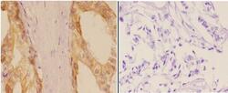
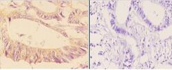
- Immunohistochemistry analysis of Phospho-ADD1/ADD2 (Ser726, Ser713) in paraffin-embedded human breast carcinoma tissue (cytoplasm and membrane staining) and negative control (right, with PBS only). Samples were incubated with Phospho-ADD1/ADD2 (Ser726, Ser713) polyclonal antibody (Product # PA5-36614) at a dilution of 1:50, followed by goat Anti-Rabbit IgG-biotin and avidin peroxidase.
- Submitted by
- Invitrogen Antibodies (provider)
- Main image
- Experimental details
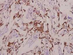
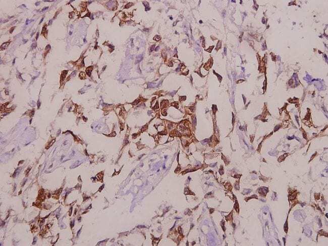
- Immunohistochemical analysis of Phospho-ADD1 pSer726+ADD2 pSer713 in paraffin-embedded human colorectal carcinoma using Phospho-ADD1 pSer726+ADD2 pSer713 polyclonal antibody (Product # PA5-36614) at a dilution of 1:50.
- Submitted by
- Invitrogen Antibodies (provider)
- Main image
- Experimental details
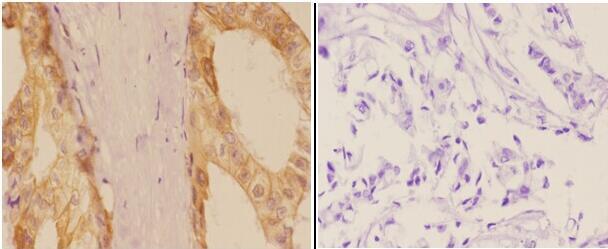
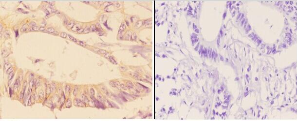
- Immunohistochemistry analysis of Phospho-ADD1/ADD2 (Ser726, Ser713) in paraffin-embedded human rectum carcinoma tissue (membrane and cytoplasmic staining) and negative control (right, with PBS only). Samples were incubated with Phospho-ADD1/ADD2 (Ser726, Ser713) polyclonal antibody (Product # PA5-36614) at a dilution of 1:50, followed by goat Anti-Rabbit IgG-biotin and avidin peroxidase.
Supportive validation
- Submitted by
- Invitrogen Antibodies (provider)
- Main image

- Experimental details
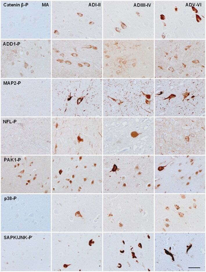
- FIGURE 7 Immunohistochemistry of selected phosphorylated proteins in the EC at different stages of NFT pathology (stages I-II, III-IV, and V-VI of Braak). Phosphorylated catenin-beta and p38-P immunoreactivity appear at the first and middle stages of NFT pathology as small granules in the cytoplasm of a subpopulation of neurons. This pattern is also found for p38-P in many neurons of the EC at stages V-VI of NFT pathology. MAP2-P, NFL-P, and SAPK/JNK-P immunoreactivity is found in neurons with the morphology of NFTs from the first stages onwards; the number of affected neurons increases with disease progression. The number of NFL-P-positive neurons is, by far, smaller than the number of neurons with MAP2-P pathology in consecutive sections. PAK1-P immunoreactivity is seen in NFTs at stages III-IV and V-VI, but as irregular or granular deposits in EC neurons at staged I-II. ADD1-P immunoreactivity increases in astrocytes and in a subpopulation of NFTs at middle and advanced stages of NFT pathology. Paraffin sections, lightly counterstained with hematoxylin, bar = 50 mum