 Explore
 Validate
 Learn
Learn Western blot
 Immunocytochemistry
Antibody data
- Antibody Data
- Antigen structure
- References [0]
- Comments [0]
- Validations
- Western blot [1]
- Immunohistochemistry [1]
- Flow cytometry [1]
Submit
Validation data
Reference
Comment
Report error
- Product number
- NBP2-59668 - Provider product page

- Provider
- Novus Biologicals
- Product name
- Rabbit Monoclonal Spectrin beta 3 Antibody
- Antibody type
- Monoclonal
- Description
- Protein A or G purified.
- Reactivity
- Human, Mouse
- Host
- Rabbit
- Isotype
- IgG
- Vial size
- 0.1 mg
- Concentration
- 1.0 mg/ml
- Storage
- Store at 4C short term. Aliquot and store at -20C long term. Avoid freeze-thaw cycles.
No comments: Submit comment
Supportive validation
- Submitted by
- Novus Biologicals (provider)
- Main image
- Experimental details
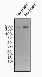
- Western Blot: Spectrin beta 3 Antibody (1287A) [NBP2-59668] - Total protein from Human and Mouse Brain was separated on a 7.5% gel by SDS-PAGE, transferred to PVDF membrane and blocked in 5% non-fat milk in TBST. The membrane was probed with 2.0 ug/ml anti-Spectrin Beta III in blocking buffer and detected with an anti-rabbit HRP secondary antibody using chemiluminescence.

Supportive validation
- Submitted by
- Novus Biologicals (provider)
- Main image

- Experimental details
- Immunohistochemistry-Paraffin: Spectrin beta 3 Antibody (1287A) [NBP2-59668] - IHC analysis of a formalin fixed paraffin embedded (FFPE) tissue section of human brain using Spectrin beta III antibody at a 1:1,000 dilution. The primary antibody bound to Spectrin beta III was detected with HRP-DAB detection method and the nuclei were counterstained with hematoxylin. This Spectrin beta III antibody generated a specific cytoplasmic staining in all the cells with strongest signal in neuronal cells.

Supportive validation
- Submitted by
- Novus Biologicals (provider)
- Main image

- Experimental details
- Flow (Intracellular): Spectrin beta 3 Antibody (1287A) [NBP2-59668] - An intracellular stain was performed on HeLa Cells with Spectrin beta 3 (1287A) antibody NBP2-59668 (blue) and a matched isotype control MAB1050 (orange). Cells were fixed with 4% paraformaldehyde, following fixation, cells were permeabilized with 0.1% saponin. Cells were incubated in an antibody dilution of 1 ug/mL for 30 minutes at room temperature, followed by rabbit IgG APC-conjugated secondary antibody (F0111, R&D Systems).
