 Explore
 Validate
 Learn
Learn Western blot
Antibody data
- Antibody Data
- Antigen structure
- References [8]
- Comments [0]
- Validations
- Western blot [1]
- Immunohistochemistry [1]
- Blocking/Neutralizing [1]
Submit
Validation data
Reference
Comment
Report error
- Product number
- AF788 - Provider product page

- Provider
- R&D Systems
- Product name
- Human/Mouse/Rat GDF-8/Myostatin Antibody
- Antibody type
- Polyclonal
- Description
- Antigen Affinity-purified. Detects human, mouse, and rat GDF-8/Myostatin in direct ELISAs and Western blots. In direct ELISAs and Western blots, approximately 30% cross-reactivity is observed with recombinant human/mouse/rat GDF-11. In Western blots, no cross-reactivity with recombinant human BMP-6 and recombinant mouse BMP-6 is observed.
- Reactivity
- Human, Mouse, Rat
- Host
- Goat
- Conjugate
- Unconjugated
- Antigen sequence
O08689- Isotype
- IgG
- Vial size
- 100 ug
- Concentration
- LYOPH
- Storage
- Use a manual defrost freezer and avoid repeated freeze-thaw cycles. 12 months from date of receipt, -20 to -70 °C as supplied. 1 month, 2 to 8 °C under sterile conditions after reconstitution. 6 months, -20 to -70 °C under sterile conditions after reconstitution.
Submitted references A GDF11/myostatin inhibitor, GDF11 propeptide-Fc, increases skeletal muscle mass and improves muscle strength in dystrophic mdx mice.
The Use of Platelet-Rich and Platelet-Poor Plasma to Enhance Differentiation of Skeletal Myoblasts: Implications for the Use of Autologous Blood Products for Muscle Regeneration.
Compensatory anabolic signaling in the sarcopenia of experimental chronic arthritis.
Myostatin signaling is up-regulated in female patients with advanced heart failure.
Inhibition of GDF8 (Myostatin) accelerates bone regeneration in diabetes mellitus type 2.
A single heterochronic blood exchange reveals rapid inhibition of multiple tissues by old blood.
High concentrations of HGF inhibit skeletal muscle satellite cell proliferation in vitro by inducing expression of myostatin: a possible mechanism for reestablishing satellite cell quiescence in vivo.
Increased secretion and expression of myostatin in skeletal muscle from extremely obese women.
Jin Q, Qiao C, Li J, Xiao B, Li J, Xiao X
Skeletal muscle 2019 May 27;9(1):16
Skeletal muscle 2019 May 27;9(1):16
The Use of Platelet-Rich and Platelet-Poor Plasma to Enhance Differentiation of Skeletal Myoblasts: Implications for the Use of Autologous Blood Products for Muscle Regeneration.
Miroshnychenko O, Chang WT, Dragoo JL
The American journal of sports medicine 2017 Mar;45(4):945-953
The American journal of sports medicine 2017 Mar;45(4):945-953
Compensatory anabolic signaling in the sarcopenia of experimental chronic arthritis.
Little RD, Prieto-Potin I, Pérez-Baos S, Villalvilla A, Gratal P, Cicuttini F, Largo R, Herrero-Beaumont G
Scientific reports 2017 Jul 24;7(1):6311
Scientific reports 2017 Jul 24;7(1):6311
Myostatin signaling is up-regulated in female patients with advanced heart failure.
Ishida J, Konishi M, Saitoh M, Anker M, Anker SD, Springer J
International journal of cardiology 2017 Jul 1;238:37-42
International journal of cardiology 2017 Jul 1;238:37-42
Inhibition of GDF8 (Myostatin) accelerates bone regeneration in diabetes mellitus type 2.
Wallner C, Jaurich H, Wagner JM, Becerikli M, Harati K, Dadras M, Lehnhardt M, Behr B
Scientific reports 2017 Aug 29;7(1):9878
Scientific reports 2017 Aug 29;7(1):9878
A single heterochronic blood exchange reveals rapid inhibition of multiple tissues by old blood.
Rebo J, Mehdipour M, Gathwala R, Causey K, Liu Y, Conboy MJ, Conboy IM
Nature communications 2016 Nov 22;7:13363
Nature communications 2016 Nov 22;7:13363
High concentrations of HGF inhibit skeletal muscle satellite cell proliferation in vitro by inducing expression of myostatin: a possible mechanism for reestablishing satellite cell quiescence in vivo.
Yamada M, Tatsumi R, Yamanouchi K, Hosoyama T, Shiratsuchi S, Sato A, Mizunoya W, Ikeuchi Y, Furuse M, Allen RE
American journal of physiology. Cell physiology 2010 Mar;298(3):C465-76
American journal of physiology. Cell physiology 2010 Mar;298(3):C465-76
Increased secretion and expression of myostatin in skeletal muscle from extremely obese women.
Hittel DS, Berggren JR, Shearer J, Boyle K, Houmard JA
Diabetes 2009 Jan;58(1):30-8
Diabetes 2009 Jan;58(1):30-8
No comments: Submit comment
Supportive validation
- Submitted by
- R&D Systems (provider)
- Main image

- Experimental details
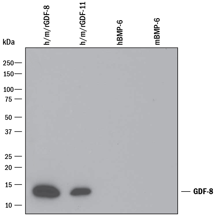
- Detection of Recombinant Human, Mouse, and Rat GDF-8/Myostatin by Western Blot. Western blot shows 25 ng of Recombinant Human/Mouse/Rat GDF-8/Myostatin (Catalog # 788-G8), Recombinant Human/Mouse/Rat GDF-11/BMP-11 (Catalog # 1958-GD), Recombinant Human BMP-6 (Catalog # 507-BP), and Recombinant Mouse BMP-6 (Catalog # 6325-BM). PVDF Membrane was probed with 0.1 µg/mL of Goat Anti-Human/Mouse/Rat GDF-8/Myostatin Antigen Affinity-purified Polyclonal Antibody (Catalog # AF788) followed by HRP-conjugated Anti-Goat IgG Secondary Antibody (Catalog # HAF109). A specific band was detected for GDF-8/Myostatin at approximately 14 kDa (as indicated). This experiment was conducted under reducing conditions and using Immunoblot Buffer Group 3.
Supportive validation
- Submitted by
- R&D Systems (provider)
- Main image

- Experimental details
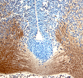
- GDF-8/Myostatin in Mouse Embryo. GDF-8/Myostatin was detected in immersion fixed frozen sections of mouse embryo (10 d.p.c., section through neural tube) using Goat Anti-Human/Mouse/Rat GDF-8/Myostatin Antigen Affinity-purified Polyclonal Antibody (Catalog # AF788) at 15 µg/mL overnight at 4 °C. Tissue was stained using the Anti-Goat HRP-DAB Cell & Tissue Staining Kit (brown; Catalog # CTS008) and counterstained with hematoxylin (blue). View our protocol for Chromogenic IHC Staining of Frozen Tissue Sections.
Supportive validation
- Submitted by
- R&D Systems (provider)
- Main image
- Experimental details
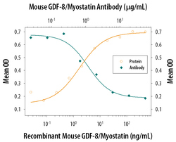
- Hemoglobin Expression Induced by GDF-8/Myostatin and Neutralization by Mouse GDF-8/Myostatin Antibody. Recombinant Mouse GDF-8/Myostatin (Catalog # 788-G8) increases hemoglobin expression in the K562 human chronic myelogenous leukemia cell line in a dose-dependent manner (orange line), as measured by the psuedoperoxidase assay. Hemoglobin expression elicited by Recombinant Mouse GDF-8/Myostatin (30 ng/mL) is neutralized (green line) by increasing concentrations of Goat Anti-Human/Mouse/Rat GDF-8/Myostatin Antigen Affinity-purified Polyclonal Antibody (Catalog # AF788). The ND50 is typically 0.6-3 µg/mL.
