 Explore
 Validate
 Learn
Learn Western blot
 Immunocytochemistry
Antibody data
- Antibody Data
- Antigen structure
- References [0]
- Comments [0]
- Validations
- Western blot [4]
- Immunohistochemistry [2]
Submit
Validation data
Reference
Comment
Report error
- Product number
- LS-C287177 - Provider product page

- Provider
- LSBio
- Product name
- TRIM28 / KAP1 Antibody (aa1-50) LS-C287177
- Antibody type
- Polyclonal
- Description
- Affinity purified
- Reactivity
- Human, Mouse
- Host
- Rabbit
- Isotype
- IgG
- Storage
- Store at 2-8°C for up to one year.
No comments: Submit comment
Enhanced validation
- Submitted by
- LSBio (provider)
- Enhanced method
- Genetic validation
- Main image
- Experimental details
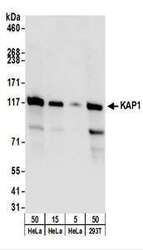
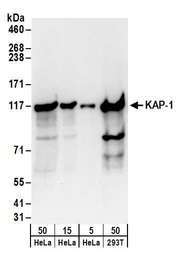
- Detection of Human KAP1 by Western Blot. Samples: Whole cell lysate from HeLa (5, 15, 50 ug), and 293T (50 ug) cells. Antibodies: Affinity purified rabbit anti-KAP1 antibody used for WB at 0.1 ug/ml. Detection: Chemiluminescence with an exposure time of 3 seconds.
- Submitted by
- LSBio (provider)
- Enhanced method
- Genetic validation
- Main image
- Experimental details
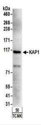
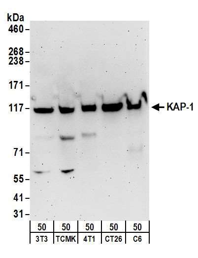
- Detection of Mouse KAP1 by Western Blot. Samples: Whole cell lysate from TCMK-1 (50 ug) cells. Antibodies: Affinity purified rabbit anti-KAP1 antibody used for WB at 1 ug/ml. Detection: Chemiluminescence with an exposure time of 3 minutes.
- Submitted by
- LSBio (provider)
- Enhanced method
- Genetic validation
- Main image
- Experimental details
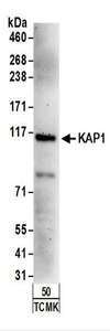
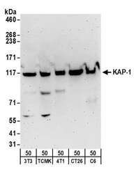
- Detection of mouse and rat KAP-1 by western blot. Samples: Whole cell lysate (50 µg) from NIH 3T3, TCMK-1, 4T1, CT26.WT, and rat C6 cells. Antibodies: Affinity purified rabbit anti-KAP-1 antibody used for WB at 1 µg/ml. Detection: Chemiluminescence with an exposure time of 3 minutes.
- Submitted by
- LSBio (provider)
- Enhanced method
- Genetic validation
- Main image

- Experimental details
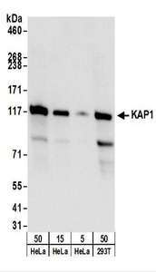
- Detection of human KAP-1 by western blot. Samples: Whole cell lysate from HeLa (5, 15 and 50 µg), and HEK293T (50µg) cells. Antibodies: Affinity purified rabbit anti-KAP-1 antibody used for WB at 0.1 µg/ml. Detection: Chemiluminescence with an exposure time of 30 seconds.
Supportive validation
- Submitted by
- LSBio (provider)
- Enhanced method
- Genetic validation
- Main image

- Experimental details
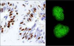
- Detection of Human KAP-1 by Immunohistochemistry and Immunocytochemistry. Sample: FFPE section of human prostate carcinoma (left) and formaldelyde-fixed HeLa cells (right). Antibody: Affinity purified rabbit anti-KAP-1 used at a dilution of 1:1000 (1μg/ml). Detection: DAB (left) and green fluorescent goat anti-rabbit IgG highly cross-adsorbed Antibody FITC conjugated used at a dilution of 1:100 (right).

- Submitted by
- LSBio (provider)
- Enhanced method
- Genetic validation
- Main image
- Experimental details
- Detection of Human KAP-1 by Immunohistochemistry and Immunocytochemistry. Sample: FFPE section of human prostate carcinoma (left) and formaldelyde-fixed HeLa cells (right). Antibody: Affinity purified rabbit anti-KAP-1 used at a dilution of 1:1000 (1μg/ml). Detection: DAB (left) and green fluorescent goat anti-rabbit IgG highly cross-adsorbed Antibody FITC conjugated used at a dilution of 1:100 (right).
