Explore
 Validate
 Learn
Learn Western blot
 Immunocytochemistry
Antibody data
- Antibody Data
- Antigen structure
- References [1]
- Comments [0]
- Validations
- Immunocytochemistry [6]
- Immunoprecipitation [1]
- Immunohistochemistry [3]
- Chromatin Immunoprecipitation [3]
- Other assay [3]
Submit
Validation data
Reference
Comment
Report error
- Product number
- PA5-27648 - Provider product page

- Provider
- Invitrogen Antibodies
- Product name
- TRIM28 Polyclonal Antibody
- Antibody type
- Polyclonal
- Antigen
- Recombinant full-length protein
- Description
- Recommended positive controls: H1299, HeLa, NIH-3T3. Predicted reactivity: Mouse (91%), Rat (91%), Bovine (93%). Store product as a concentrated solution. Centrifuge briefly prior to opening the vial.
- Reactivity
- Human, Mouse, Rat
- Host
- Rabbit
- Isotype
- IgG
- Vial size
- 100 μL
- Concentration
- 0.48 mg/mL
- Storage
- Store at 4°C short term. For long term storage, store at -20°C, avoiding freeze/thaw cycles.
Submitted references The Anti-Glucocorticoid Receptor Antibody Clone 5E4: Raising Awareness of Unspecific Antibody Binding.
Ehlers L, Kirchner M, Mertins P, Strehl C, Buttgereit F, Gaber T
International journal of molecular sciences 2022 May 2;23(9)
International journal of molecular sciences 2022 May 2;23(9)
No comments: Submit comment
Supportive validation
- Submitted by
- Invitrogen Antibodies (provider)
- Main image

- Experimental details
- Immunofluorescent analysis of KAP1 in paraformaldehyde-fixed A431 cells using a KAP1 polyclonal antibody (Product # PA5-27648) at a 1:200 dilution.
- Submitted by
- Invitrogen Antibodies (provider)
- Main image

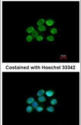
- Experimental details
- Immunocytochemistry-Immunofluorescence analysis of TRIM28 was performed in HeLa cells fixed in 4% paraformaldehyde at RT for 15 min. Green: TRIM28 Polyclonal Antibody (Product # PA5-27648) diluted at 1:200. Red: alpha Tubulin, a cytoskeleton marker.

- Submitted by
- Invitrogen Antibodies (provider)
- Main image
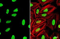
- Experimental details
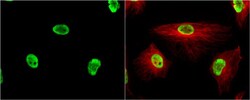
- TRIM28 Polyclonal Antibody detects KAP1 protein at nucleus by immunofluorescent analysis. Sample: HeLa cells were fixed in 4% paraformaldehyde at RT for 15 min. Green: KAP1 stained by TRIM28 Polyclonal Antibody (Product # PA5-27648) diluted at 1:2,000. Red: phalloidin, a cytoskeleton marker, diluted at 1:200. Scale bar= 10 µm.

- Submitted by
- Invitrogen Antibodies (provider)
- Main image
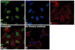
- Experimental details
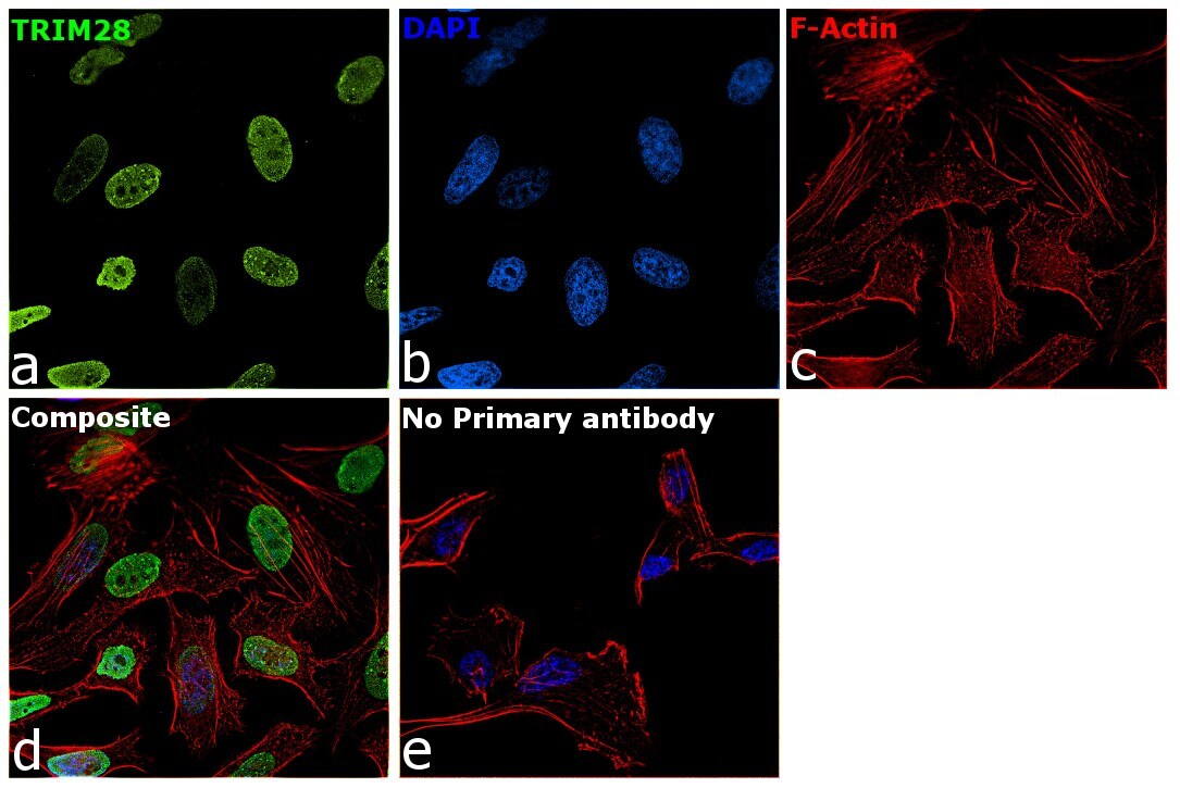
- Immunofluorescence analysis of TRIM28 was performed using 70 confluent log phase HeLa cells. The cells were fixed with 4% paraformaldehyde for 10 minutes, permeabilized with 0.1% Triton™ X-100 for 15 minutes, and blocked with 2% BSA for 45 minutes at room temperature. The cells were labeled with TRIM28 Polyclonal Antibody (Product # PA5-27648) at 1:100 dilution in 0.1% BSA, incubated at 4 degree celsius overnight and then labeled with Donkey anti-Rabbit IgG (H+L) Highly Cross-Adsorbed Secondary Antibody, Alexa Fluor Plus 488 (Product # A32790), (1:2000 dilution), for 45 minutes at room temperature (Panel a: Green). Nuclei (Panel b:Blue) were stained with ProLong™ Diamond Antifade Mountant with DAPI (Product # P36962). F-actin (Panel c: Red) was stained with Rhodamine Phalloidin (Product # R415, 1:300 dilution). Panel d represents the merged image showing Nuclear localization. Panel e represents control cells with no primary antibody to assess background. The images were captured at 60X magnification.
- Submitted by
- Invitrogen Antibodies (provider)
- Main image
- Experimental details
- Immunocytochemistry-Immunofluorescence analysis of TRIM28 was performed in HeLa cells fixed in 4% paraformaldehyde at RT for 15 min. Green: TRIM28 Polyclonal Antibody (Product # PA5-27648) diluted at 1:200. Red: alpha Tubulin, a cytoskeleton marker.

- Submitted by
- Invitrogen Antibodies (provider)
- Main image

- Experimental details
- Immunofluorescence analysis of TRIM28 was performed using 70 confluent log phase HeLa cells. The cells were fixed with 4% paraformaldehyde for 10 minutes, permeabilized with 0.1% Triton™ X-100 for 15 minutes, and blocked with 2% BSA for 45 minutes at room temperature. The cells were labeled with TRIM28 Polyclonal Antibody (Product # PA5-27648) at 1:100 dilution in 0.1% BSA, incubated at 4 degree celsius overnight and then labeled with Donkey anti-Rabbit IgG (H+L) Highly Cross-Adsorbed Secondary Antibody, Alexa Fluor Plus 488 (Product # A32790), (1:2000 dilution), for 45 minutes at room temperature (Panel a: Green). Nuclei (Panel b:Blue) were stained with ProLong™ Diamond Antifade Mountant with DAPI (Product # P36962). F-actin (Panel c: Red) was stained with Rhodamine Phalloidin (Product # R415, 1:300 dilution). Panel d represents the merged image showing Nuclear localization. Panel e represents control cells with no primary antibody to assess background. The images were captured at 60X magnification.

Supportive validation
- Submitted by
- Invitrogen Antibodies (provider)
- Main image

- Experimental details
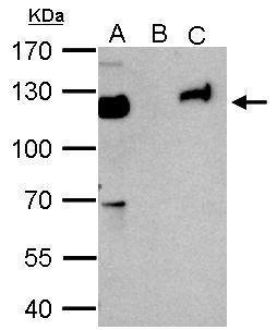
- KAP1 antibody immunoprecipitates KAP1 protein in IP experiments. IP Sample: 1,000 µg HeLa whole cell lysate/extract A. 50 µg HeLa whole cell lysate/extract B. Control with 2 µg of preimmune rabbit IgG C. Immunoprecipitation of KAP1 protein by 2 µg of KAP1 antibody (Product # PA5-27648) 7.5% SDS-PAGE The immunoprecipitated KAP1 protein was detected by KAP1 antibody (Product # PA5-27648) diluted at 1:1,000.

Supportive validation
- Submitted by
- Invitrogen Antibodies (provider)
- Main image
- Experimental details
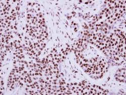
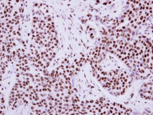
- Immunohistochemical analysis of paraffin-embedded human breast cancer, using KAP1 (Product # PA5-27648) antibody at 1:500 dilution. Antigen Retrieval: EDTA based buffer, pH 8.0, 15 min.
- Submitted by
- Invitrogen Antibodies (provider)
- Main image
- Experimental details
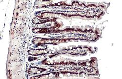
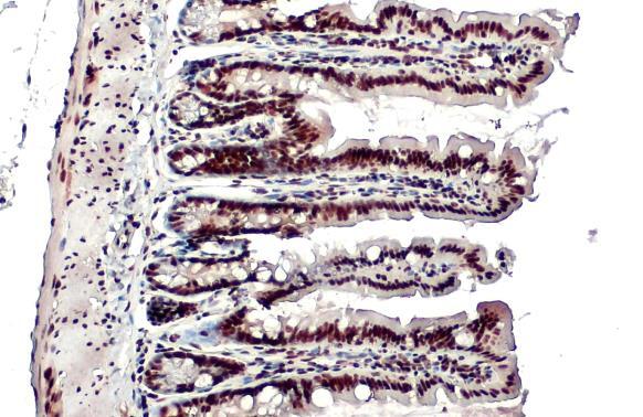
- TRIM28 Polyclonal Antibody detects KAP1 protein at nucleus by immunohistochemical analysis. Sample: Paraffin-embedded mouse intestine. KAP1 stained by TRIM28 Polyclonal Antibody (Product # PA5-27648) diluted at 1:500. Antigen Retrieval: Citrate buffer, pH 6.0, 15 min.
- Submitted by
- Invitrogen Antibodies (provider)
- Main image
- Experimental details
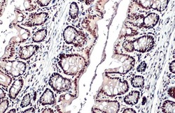
- TRIM28 Polyclonal Antibody detects KAP1 protein at nucleus by immunohistochemical analysis. Sample: Paraffin-embedded mouse colon. KAP1 stained by TRIM28 Polyclonal Antibody (Product # PA5-27648) diluted at 1:500. Antigen Retrieval: Citrate buffer, pH 6.0, 15 min.

Supportive validation
- Submitted by
- Invitrogen Antibodies (provider)
- Main image
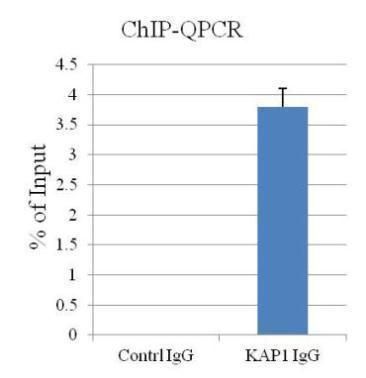
- Experimental details
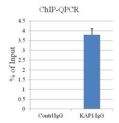
- ChIP assay followed by QPCR on a known KAP1-binding region within the ZNF180 3’UTR using a KAP1 polyclonal antibody (Product # PA5-27648).
- Submitted by
- Invitrogen Antibodies (provider)
- Main image

- Experimental details
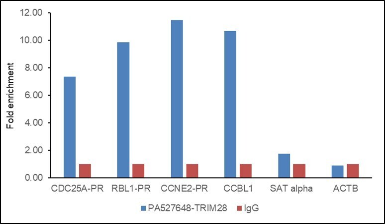
- Chromatin Immunoprecipitation (ChIP) assay of endogenous TRIM28 protein using TRIM28 Antibody: ChIP was performed using TRIM28 Polyclonal Antibody (Product # PA5-27648, 5 µg) on sheared chromatin from HeLa cells using the MAGnify ChIP System kit (Product # 49-2024). Normal Rabbit IgG was used as a negative IP control. The purified DNA was analyzed by qPCR using primers binding to CDC25- Promoter, RBL1-Promoter, CCNE2- Promoter and CCBL1 (active) and SAT alpha satellite repeats and Actin beta (Inactive). Data is presented as fold enrichment of the antibody signal versus the negative control IgG using the comparative CT method.
- Submitted by
- Invitrogen Antibodies (provider)
- Main image
- Experimental details
- Chromatin Immunoprecipitation (ChIP) assay of endogenous TRIM28 protein using TRIM28 Antibody: ChIP was performed using TRIM28 Polyclonal Antibody (Product # PA5-27648, 5 µg) on sheared chromatin from HeLa cells using the MAGnify ChIP System kit (Product # 49-2024). Normal Rabbit IgG was used as a negative IP control. The purified DNA was analyzed by qPCR using primers binding to CDC25- Promoter, RBL1-Promoter, CCNE2- Promoter and CCBL1 (active) and SAT alpha satellite repeats and Actin beta (Inactive). Data is presented as fold enrichment of the antibody signal versus the negative control IgG using the comparative CT method.

Supportive validation
- Submitted by
- Invitrogen Antibodies (provider)
- Main image

- Experimental details
- KAP1 antibody immunoprecipitates KAP1 protein in IP experiments. IP Sample: 1,000 µg HeLa whole cell lysate/extract A. 50 µg HeLa whole cell lysate/extract B. Control with 2 µg of preimmune rabbit IgG C. Immunoprecipitation of KAP1 protein by 2 µg of KAP1 antibody (Product # PA5-27648) 7.5% SDS-PAGE The immunoprecipitated KAP1 protein was detected by KAP1 antibody (Product # PA5-27648) diluted at 1:1,000.
- Submitted by
- Invitrogen Antibodies (provider)
- Main image
- Experimental details
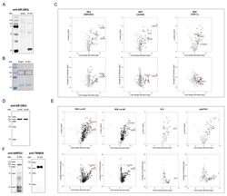
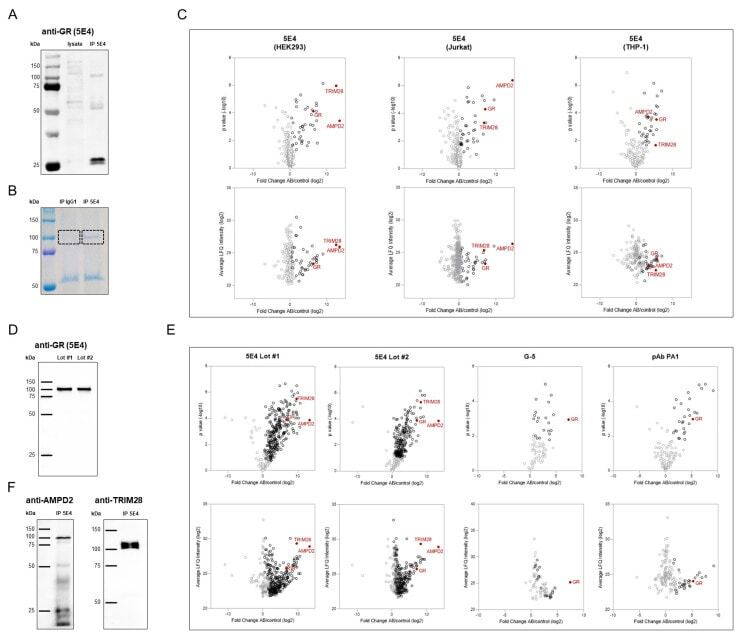
- Verification of anti-GR (5E4) antibody specificity. ( A ) Western blot analysis of GR pulled down from HEK293 membrane fractions by immunoprecipitation using the anti-GR (5E4) antibody (IP 5E4). An amount of 20 uL of membrane fraction protein (lysate) were analyzed in parallel. Protein detection was achieved by adding the anti-GR (5E4) antibody followed by an HRP-conjugated anti-mouse IgG antibody as a secondary reagent. ( B ) Immunoprecipitation from HEK293 membrane fractions was performed using the anti-GR (5E4) antibody (IP 5E4) and mouse IgG1 as a corresponding isotype control (IP IgG1). For mass spectrometric analysis the protein content was visualized by Pierce Coomassie Brilliant Blue G-250 Dye after SDS-PAGE, and the indicated area of interest was extracted for analysis. ( C ) Mass spectrometric analyses of pull-down samples obtained by immunoprecipitation from HEK293, Jurkat, and THP-1 whole cell lysates using the anti-GR (5E4) antibody (AB) and mouse IgG1 as corresponding isotype control (Control). Differential protein abundance compared to isotype control was determined using a two-sample Student's t test and black circles represent significance with an FDR cut-off of 5%. ( D ) Western blot analysis of GR pulled down from HEK293 whole cell lysates by immunoprecipitation using anti-GR (5E4) antibody Lot #1 (provided by Timea Berki []) and Lot #2 (Bio-Rad, Cat# MCA2469, RRID:AB_10844347). The protein content was visualized by incubation with anti-GR (5E4) antibody fo
- Submitted by
- Invitrogen Antibodies (provider)
- Main image
- Experimental details
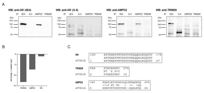
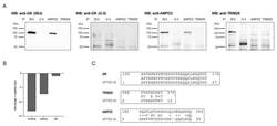
- Re-evaluation of anti-GR (5E4) antibody specificity. ( A ) Comparison of target proteins by western blot analysis. Pull-down samples from HEK293 whole cell lysates were obtained by immunoprecipitation using the following antibodies: anti-GR (5E4), anti-GR (G-5), anti-AMPD2 (QQ13), and anti-TRIM28 (Cat# PA5-27648). The protein content was visualized by incubation with primary antibodies directed against GR (5E4, biotinylated), GR (G-5, biotinylated), AMPD2 (PA5-26127, biotinylated), and TRIM28 (Cat# PA5-27648) as indicated, followed by HRP-conjugated streptavidin and anti-rabbit IgG antibody as secondary reagents. ( B ) Mass spectrometric analysis of pull-down samples obtained by IP from HEK293 whole cell lysates using the anti-GR antibody, 5E4, with and without prior two-hour incubation with APTEK-26 peptide. Bar graphs show fold change of peptide incubation to without peptide incubation. ( C ) Amino acid sequences of the newly identified anti-GR (5E4) target proteins, AMPD2 (UniProt ID: Q01433) and TRIM28 (UniProt ID: Q13263), were blasted against the APTEK-26 peptide using NCBI Protein BLAST ( https://blast.ncbi.nlm.nih.gov/Blast.cgi , accessed on 14 November 2020) [].