Explore
 Validate
 Learn
Learn Western blot
Antibody data
- Antibody Data
- Antigen structure
- References [0]
- Comments [0]
- Validations
- Western blot [1]
- Immunocytochemistry [1]
- Immunoprecipitation [1]
- Flow cytometry [1]
Submit
Validation data
Reference
Comment
Report error
- Product number
- AM33115PU-N - Provider product page

- Provider
- Acris Antibodies GmbH
- Product name
- anti EDG-1 / S1PR1
- Antibody type
- Monoclonal
- Antigen
- Other
- Reactivity
- Human
- Host
- Mouse
- Isotype
- IgG
- Antibody clone number
- 2B9
- Vial size
- 0.1 mg
- Concentration
- 1 mg/ml
No comments: Submit comment
Supportive validation
- Submitted by
- Acris Antibodies GmbH (provider)
- Main image

- Experimental details
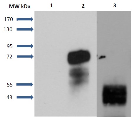
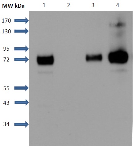
- Figure 1. Detection of S1P1 receptors in transfected cells and human umbilical vein endothelial cells (HUVEC): Lane 1: CHO-K1 cell membranes. Lane 2: S1P1-GFP CHO cell membranes. Lane 3: HUVEC membranes. CHO-K1 cells were stably transfected with S1P1-GFP construct (cell clone was a generous gift from Kevin R, Lynch). Samples were analysed by SDS-PAGE and immunoblotted with Mouse Anti-S1PR1 Monoclonal Antibody (clone 2B9) (1/5000 w/v dilution). S1P1 receptor was fused with the green fluorescent protein (GFP) at its C-terminus. Molecular weights: GFP: 27 kDa, S1P1: 43 kDa, S1P1-GFP: 70 kDa.
Supportive validation
- Submitted by
- Acris Antibodies GmbH (provider)
- Main image

- Experimental details
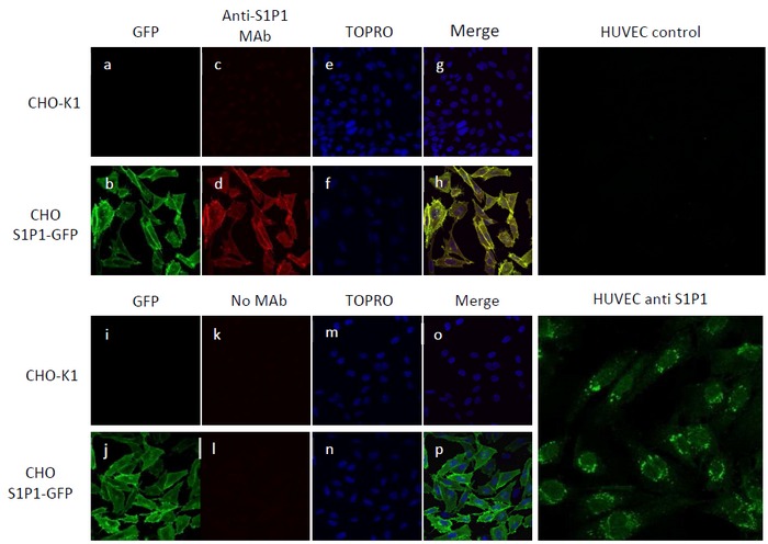
- Figure 3. Immunofluorescence detection of S1P1 receptors in transfected cells and HUVEC.Endogenous S1P1 receptors were detected by confocal microscopy in HUVEC and in transfected CHO cells stably expressing S1P1-GFP with an anti-S1P1 monoclonal antibody (1/100, w/v).CHO-K1 cells were used as negative control. S1P1-GFP receptors were detected by fluorescence (excitation 488 nm, emission 500-550nm) (a, b, i, j, green fluorescence) and immunofluorescence (Cy3-Ac II anti-mouse, excitation 559 nm, emission 570-625 nm) with ananti-S1P1 monoclonal antibody (c, d, k, l, red fluorescence). Nuclei were stained with TO-PRO-3 (excitation 635 nm, emission 655-755nm, e, f, m, and n blue fluorescence). Merge images are presented (g, h, o, p).HUVEC were stained with anti-S1P1 primary antibody and Alexa-488 secondary antibody (excitation 488 nm, emission 500-550 nm).HUVEC control was realized with omitting anti-S1P1 primary antibody.
Supportive validation
- Submitted by
- Acris Antibodies GmbH (provider)
- Main image
- Experimental details
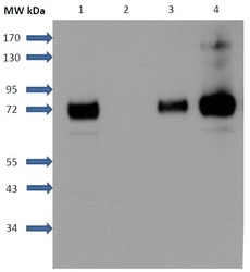
- Figure 2. Immunoprecipitation of S1P1 receptors: S1P1-GFP receptors were immunoprecipitated with a mouse monoclonal antibody anti-S1P1 after solublization of membranes with NP40 detergent and interaction with sepharose-protein G phase. Detection of precipitated receptors was realized with a rabbit secondary anti GFP antibody coupled with HRP. Lane 1: CHO-S1P1-GFP membranes. Lane 2: Control without anti-S1P1 antibodies. Lane 3: CHO-S1P1-GFP cell membranes solubilized with NP-40. Lane 4: Immunoprecipitated sample. For control and Immunoprecipitated sample, sepharose-protein G was boiled in Laemmli buffer for 5 min at 100°C.
Supportive validation
- Submitted by
- Acris Antibodies GmbH (provider)
- Main image
- Experimental details
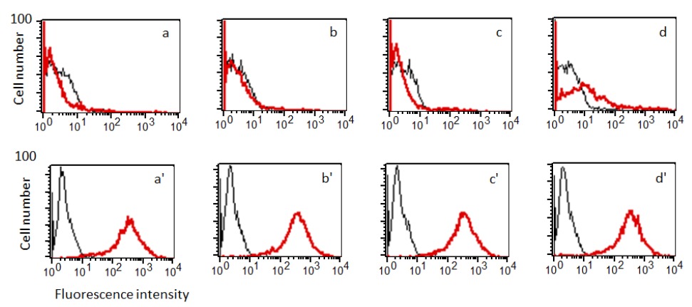
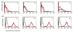
- Figure 4. Comparative evaluation of different Anti-S1P1 antibodies in Flow Cytometry: S1P1 receptors were detected with anti-S1P1 antibodies (1/100) by flow cytometry in recombinant CHO cells stably expressing S1P1.GFP (red line) and in CHO-K1 cells (black line) as negative controls. S1P1-GFP receptors were detected by fluorescence (excitation 488 nm, emission 530/30 nm) (aâ, bâ, câ, dâ) and immunofluorescence. (APC, excitation 635 nm, emission 661/16 nm) with different anti-S1P1 antibodies (a- Mab1, a-Pab1, c-Pab2 and d-Cat.-No AM33115PU).