Explore
 Validate
 Learn
Learn Western blot
Antibody data
- Antibody Data
- Antigen structure
- References [0]
- Comments [0]
- Validations
- Western blot [1]
- Immunocytochemistry [1]
- Immunohistochemistry [1]
Submit
Validation data
Reference
Comment
Report error
- Product number
- TA328613 - Provider product page

- Provider
- OriGene
- Product name
- Rabbit Polyclonal Anti-Sphingosine 1-Phosphate Receptor 1 (extracellular)
- Antibody type
- Polyclonal
- Description
- Rabbit Polyclonal Anti-Sphingosine 1-Phosphate Receptor 1 (extracellular)
- Host
- Rabbit
- Conjugate
- Unconjugated
- Epitope
- S1PR1
- Antibody clone number
- NULL
- Vial size
- 200 µl
- Concentration
- NULL
No comments: Submit comment
Supportive validation
- Submitted by
- OriGene (provider)
- Main image
- Experimental details
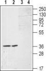
- Western blot analysis of RAEC cells (lanes 1 and 3) and in A-10 (lane 2 and 4) cell lysates: 1, 2. Anti-Sphingosine 1-Phosphate Receptor 1 (extracellular) antibody (1:200). 3, 4. Anti-Sphingosine 1-Phosphate Receptor 1 (extracellular) antibody, preincubated with the control peptide antigen.
- Validation comment
- WB

Supportive validation
- Submitted by
- OriGene (provider)
- Main image

- Experimental details
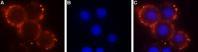
- Expression of Sphingosine 1-Phosphate Receptor 1 in mouse 3T3 cells. Immunocytochemical staining of mouse 3T3 cells using Anti-Sphingosine 1-Phosphate Receptor 1 (extracellular) antibody (1:50), (red). DAPI is used for nuclear staining (blue).
- Validation comment
- IF
Supportive validation
- Submitted by
- OriGene (provider)
- Main image
- Experimental details
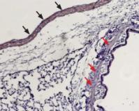
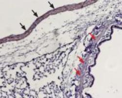
- Expression of Sphingosine 1-Phosphate Receptor 1 in rat lung. Immunohistochemical staining of paraffin embedded rat lung sections using Anti-Sphingosine 1-Phosphate Receptor 1 (extracellular) antibody(1:100). Staining is present in vascular smooth muscle (black arrows) but not in the muscular layer of bronchi (red arrows). Hematoxilin is used as the counterstain.
- Validation comment
- IHC