Explore
 Validate
 Learn
Learn Flow cytometry
Antibody data
- Antibody Data
- Antigen structure
- References [5]
- Comments [0]
- Validations
- Flow cytometry [2]
Submit
Validation data
Reference
Comment
Report error
- Product number
- FAB7089A-100UG - Provider product page

- Provider
- R&D Systems
- Product name
- Mouse S1P1/EDG-1 APC-conjugated Antibody
- Antibody type
- Monoclonal
- Description
- Protein A or G purified from hybridoma culture supernatant. Detects mouse S1P1/EDG-1 peptide in direct ELISAs.
- Reactivity
- Mouse
- Host
- Rat
- Conjugate
- Red dye
- Antigen sequence
O08530- Isotype
- IgG
- Antibody clone number
- 713412
- Vial size
- 100 ug
- Storage
- Protect from light. Do not freeze. 12 months from date of receipt, 2 to 8 °C as supplied.
Submitted references Sequestration of T cells in bone marrow in the setting of glioblastoma and other intracranial tumors.
Impaired lymphocyte trafficking in mice deficient in the kinase activity of PKN1.
Targeting of Ly9 (CD229) Disrupts Marginal Zone and B1 B Cell Homeostasis and Antibody Responses.
IL4RA on lymphatic endothelial cells promotes T cell egress during sclerodermatous graft versus host disease.
Intravenous gammaglobulin inhibits encephalitogenic potential of pathogenic T cells and interferes with their trafficking to the central nervous system, implicating sphingosine-1 phosphate receptor 1-mammalian target of rapamycin axis.
Chongsathidkiet P, Jackson C, Koyama S, Loebel F, Cui X, Farber SH, Woroniecka K, Elsamadicy AA, Dechant CA, Kemeny HR, Sanchez-Perez L, Cheema TA, Souders NC, Herndon JE, Coumans JV, Everitt JI, Nahed BV, Sampson JH, Gunn MD, Martuza RL, Dranoff G, Curry WT, Fecci PE
Nature medicine 2018 Sep;24(9):1459-1468
Nature medicine 2018 Sep;24(9):1459-1468
Impaired lymphocyte trafficking in mice deficient in the kinase activity of PKN1.
Mashud R, Nomachi A, Hayakawa A, Kubouchi K, Danno S, Hirata T, Matsuo K, Nakayama T, Satoh R, Sugiura R, Abe M, Sakimura K, Wakana S, Ohsaki H, Kamoshida S, Mukai H
Scientific reports 2017 Aug 9;7(1):7663
Scientific reports 2017 Aug 9;7(1):7663
Targeting of Ly9 (CD229) Disrupts Marginal Zone and B1 B Cell Homeostasis and Antibody Responses.
Cuenca M, Romero X, Sintes J, Terhorst C, Engel P
Journal of immunology (Baltimore, Md. : 1950) 2016 Jan 15;196(2):726-37
Journal of immunology (Baltimore, Md. : 1950) 2016 Jan 15;196(2):726-37
IL4RA on lymphatic endothelial cells promotes T cell egress during sclerodermatous graft versus host disease.
Urso K, Alvarez D, Cremasco V, Tsang K, Grauel A, Lafyatis R, von Andrian UH, Ermann J, Aliprantis AO
JCI insight 2016 Aug 4;1(12)
JCI insight 2016 Aug 4;1(12)
Intravenous gammaglobulin inhibits encephalitogenic potential of pathogenic T cells and interferes with their trafficking to the central nervous system, implicating sphingosine-1 phosphate receptor 1-mammalian target of rapamycin axis.
Othy S, Hegde P, Topçu S, Sharma M, Maddur MS, Lacroix-Desmazes S, Bayry J, Kaveri SV
Journal of immunology (Baltimore, Md. : 1950) 2013 May 1;190(9):4535-41
Journal of immunology (Baltimore, Md. : 1950) 2013 May 1;190(9):4535-41
No comments: Submit comment
Supportive validation
- Submitted by
- R&D Systems (provider)
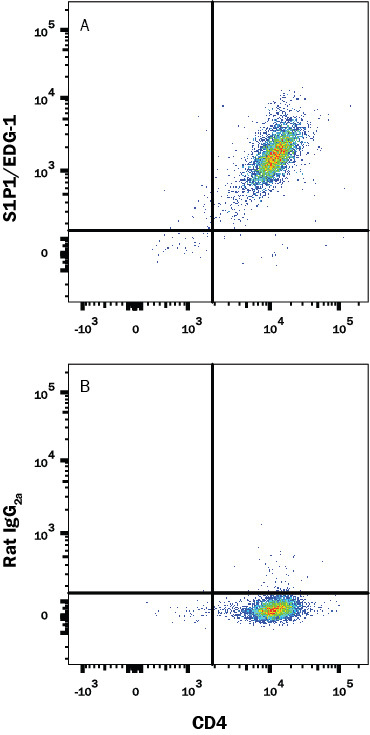
- Main image
- Experimental details
- Detection of S1P1/EDG-1 in Mouse Thymocytes by Flow Cytometry. Mouse thymocytes were stained with Rat Anti-Mouse CD4 PE-conjugated Monoclonal Antibody (Catalog # FAB554P) and either (A) Rat Anti-Mouse S1P1/EDG-1 APC-conjugated Monoclonal Antibody (Catalog # FAB7089A) or (B) Rat IgG2A Allophycocyanin Isotype Control (Catalog # IC006A). View our protocol for Staining Membrane-associated Proteins.
- Submitted by
- R&D Systems (provider)
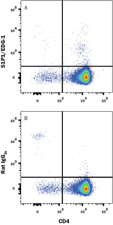
- Main image

- Experimental details
- Detection of S1P1/EDG-1 in HEK293 Human Cell Line Transfected with Mouse S1P1/EDG-1 and eGFP by Flow Cytometry. HEK293 human embryonic kidney cell line transfected with mouse S1P1/EDG-1 and eGFP was stained with either (A) Rat Anti-Mouse S1P1/EDG-1 APC-conjugated Monoclonal Antibody (Catalog # FAB7089A) or (B) Rat IgG2A Allophycocyanin Isotype Control (Catalog # IC006A). View our protocol for Staining Membrane-associated Proteins.