 Explore
 Validate
 Learn
Learn Western blot
 Immunocytochemistry
Antibody data
- Antibody Data
- Antigen structure
- References [0]
- Comments [0]
- Validations
- Immunocytochemistry [10]
- Immunohistochemistry [4]
- Flow cytometry [2]
Submit
Validation data
Reference
Comment
Report error
- Product number
- MA5-41253 - Provider product page

- Provider
- Invitrogen Antibodies
- Product name
- CRM1 Recombinant Rabbit Monoclonal Antibody (JB35-22)
- Antibody type
- Monoclonal
- Antigen
- Recombinant full-length protein
- Reactivity
- Human, Mouse, Rat
- Host
- Rabbit
- Isotype
- IgG
- Antibody clone number
- JB35-22
- Vial size
- 100 μL
- Concentration
- 1 mg/mL
- Storage
- Store at 4°C short term. For long term storage, store at -20°C, avoiding freeze/thaw cycles.
No comments: Submit comment
Supportive validation
- Submitted by
- Invitrogen Antibodies (provider)
- Main image

- Experimental details
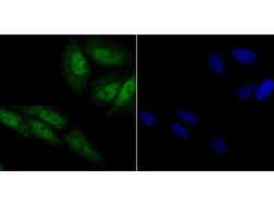
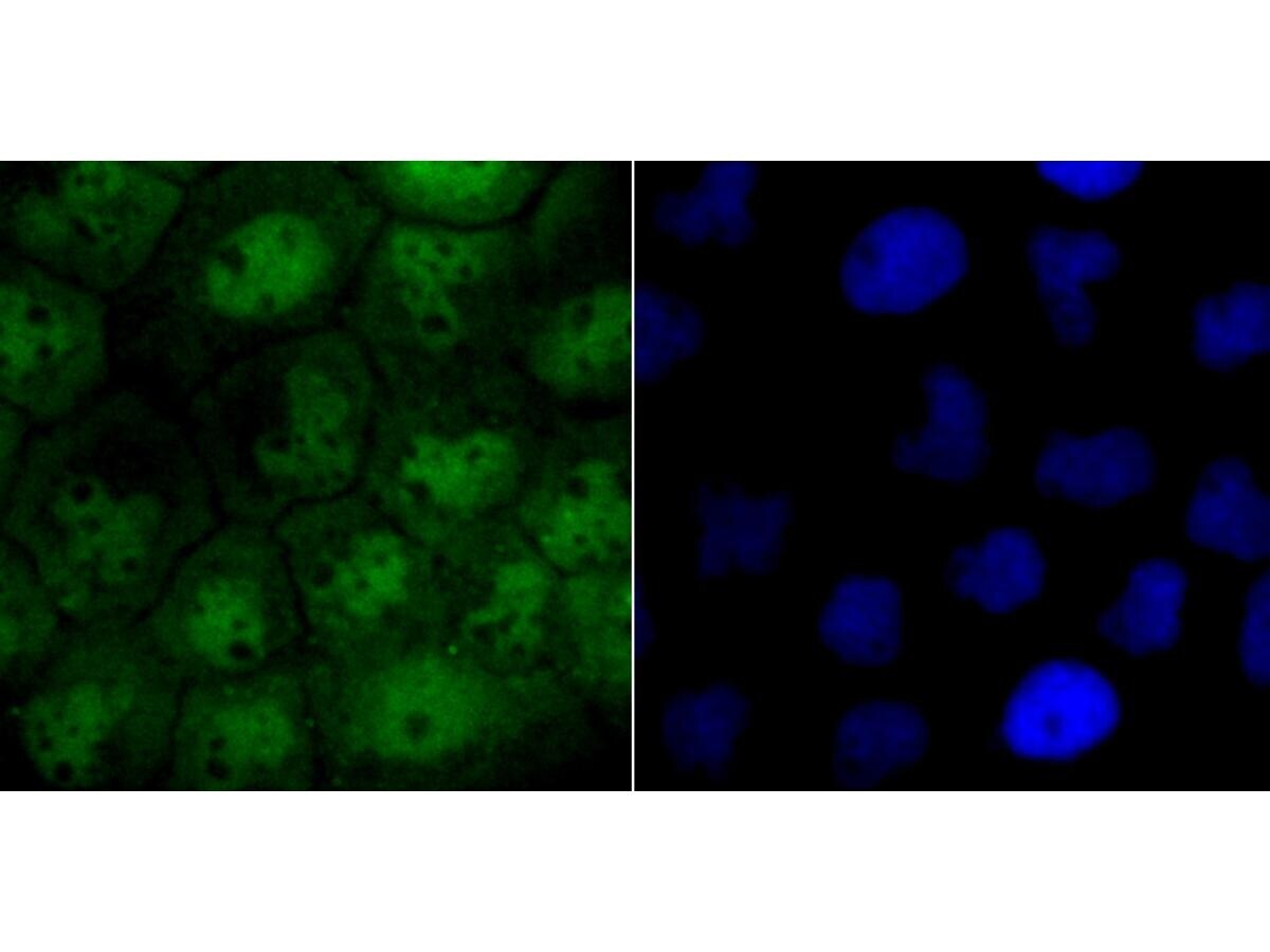
- Immunocytochemistry/Immunofluorescence analysis of CRM1 in LOVO cells (green) using CRM1 Recombinant Monoclonal Antibody (Product # MA5-41253). Cells were fixed in paraformaldehyde, permeabilised with 0.25% Triton X100/PBS. The nuclear counter stain is DAPI (blue).

- Submitted by
- Invitrogen Antibodies (provider)
- Main image
- Experimental details
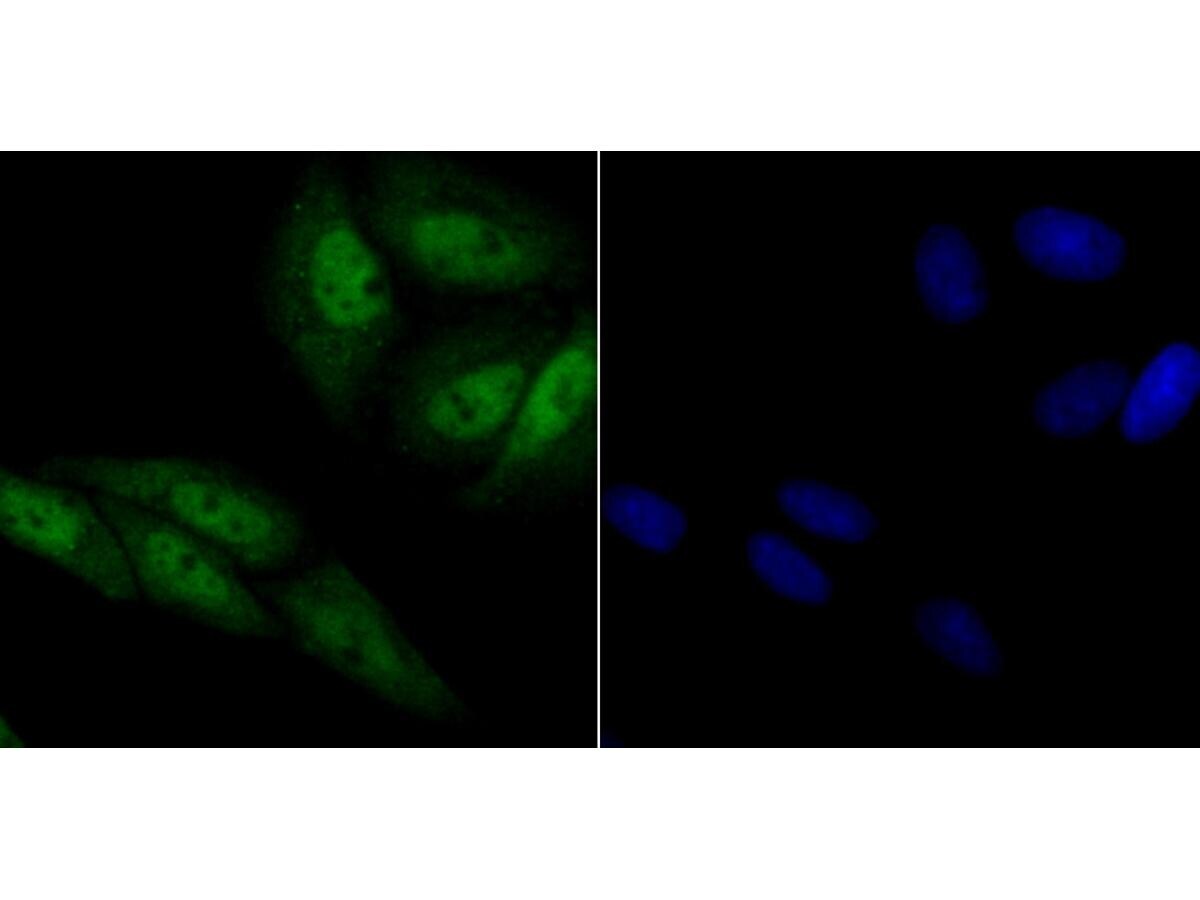
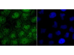
- Immunocytochemistry/Immunofluorescence analysis of CRM1 in SiHa cells (green) using CRM1 Recombinant Monoclonal Antibody (Product # MA5-41253). Cells were fixed in paraformaldehyde, permeabilised with 0.25% Triton X100/PBS. The nuclear counter stain is DAPI (blue).
- Submitted by
- Invitrogen Antibodies (provider)
- Main image

- Experimental details
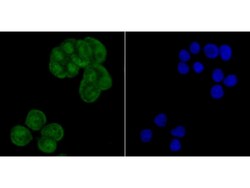
- Immunocytochemistry/Immunofluorescence analysis of CRM1 in A431 cells (green) using CRM1 Recombinant Monoclonal Antibody (Product # MA5-41253). Cells were fixed in paraformaldehyde, permeabilised with 0.25% Triton X100/PBS. The nuclear counter stain is DAPI (blue).

- Submitted by
- Invitrogen Antibodies (provider)
- Main image
- Experimental details
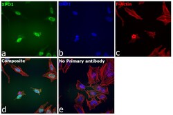
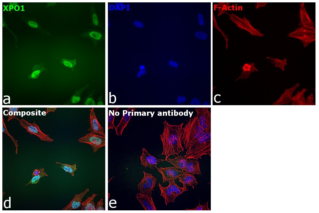
- Immunofluorescence analysis of XPO1 was performed using 70% confluent log phase HeLa cells. The cells were fixed with 4% paraformaldehyde for 15 minutes, permeabilized with 0.1% Triton™ X-100 for 15 minutes, and blocked with 2% BSA for 45 minutes at room temperature. The cells were labeled with CRM1 Recombinant Rabbit Monoclonal Antibody (JB35-22) (Product # MA5-41253) at 1:100 dilution in 0.1% BSA, incubated at 4 degree celsius overnight and then labeled with Donkey anti-Rabbit IgG (H+L) Highly Cross-Adsorbed Secondary Antibody, Alexa Fluor Plus 488 (Product # A32790), (1:2000 dilution), for 45 minutes at room temperature (Panel a: Green). Nuclei (Panel b:Blue) were stained with ProLong™ Diamond Antifade Mountant with DAPI (Product # P36962). F-actin (Panel c: Red) was stained with Rhodamine Phalloidin (Product # R415, 1:300 dilution). Panel d represents the merged image showing nuclear localization. Panel e represents control cells with no primary antibody to assess background. The images were captured at 40X magnification in CellInsight CX7 LZR High-Content Screening (HCS) Platform (Product # CX7A1110LZR) and externally deconvoluted (D.Sage et al. / Methods 115 (2017) 28–41).
- Submitted by
- Invitrogen Antibodies (provider)
- Main image

- Experimental details
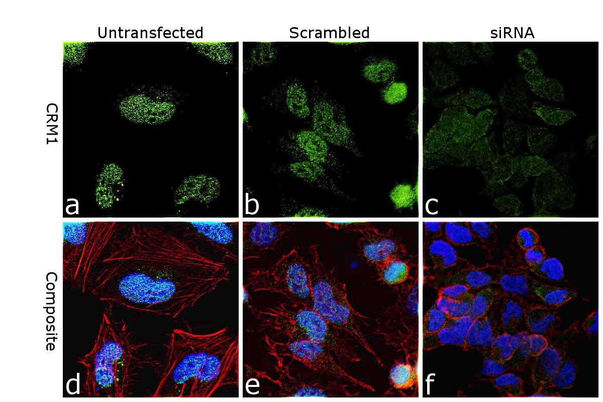
- Knockdown of XPO1 was achieved by transfecting HeLa cells with XPO1 specific siRNA (Silencer® select Product # s14937, S1439). Immunofluorescence analysis was performed on untransfected HeLa cells (panel a,d), transfected with non-specific scrambled siRNA (panels b,e) and transfected with XPO1 specific siRNA (panel c,f). Cells were fixed, permeabilized, and labelled with CRM1 Recombinant Rabbit Monoclonal Antibody (JB35-22) (Product # MA5-41253, 1:100 dilution) followed by Goat anti-Rabbit IgG (H+L) Superclonal™ Recombinant Secondary Antibody, Alexa Fluor® 488 conjugate (Product # A27034), (1:2000 dilution). Nuclei (blue) were stained using ProLong™ Diamond Antifade Mountant with DAPI (Product # P36962), and Rhodamine Phalloidin (Product # R415, 1:300 dilution) was used for cytoskeletal F-actin (Red) staining. Knockdown of specific signal was observed upon siRNA mediated knockdown (panel c,f) confirming specificity of the antibody to XPO1 (Green). The Images were captured at 60X magnification.

- Submitted by
- Invitrogen Antibodies (provider)
- Main image
- Experimental details
- Immunocytochemistry/Immunofluorescence analysis of CRM1 in LOVO cells (green) using CRM1 Recombinant Monoclonal Antibody (Product # MA5-41253). Cells were fixed in paraformaldehyde, permeabilised with 0.25% Triton X100/PBS. The nuclear counter stain is DAPI (blue).

- Submitted by
- Invitrogen Antibodies (provider)
- Main image

- Experimental details
- Immunocytochemistry/Immunofluorescence analysis of CRM1 in SiHa cells (green) using CRM1 Recombinant Monoclonal Antibody (Product # MA5-41253). Cells were fixed in paraformaldehyde, permeabilised with 0.25% Triton X100/PBS. The nuclear counter stain is DAPI (blue).

- Submitted by
- Invitrogen Antibodies (provider)
- Main image
- Experimental details
- Immunocytochemistry/Immunofluorescence analysis of CRM1 in A431 cells (green) using CRM1 Recombinant Monoclonal Antibody (Product # MA5-41253). Cells were fixed in paraformaldehyde, permeabilised with 0.25% Triton X100/PBS. The nuclear counter stain is DAPI (blue).
- Submitted by
- Invitrogen Antibodies (provider)
- Main image

- Experimental details
- Knockdown of XPO1 was achieved by transfecting HeLa cells with XPO1 specific siRNA (Silencer® select Product # s14937, S1439). Immunofluorescence analysis was performed on untransfected HeLa cells (panel a,d), transfected with non-specific scrambled siRNA (panels b,e) and transfected with XPO1 specific siRNA (panel c,f). Cells were fixed, permeabilized, and labelled with CRM1 Recombinant Rabbit Monoclonal Antibody (JB35-22) (Product # MA5-41253, 1:100 dilution) followed by Goat anti-Rabbit IgG (Heavy Chain) Superclonal™ Recombinant Secondary Antibody, Alexa Fluor® 488 conjugate (Product # A27034), (1:2000 dilution). Nuclei (blue) were stained using ProLong™ Diamond Antifade Mountant with DAPI (Product # P36962), and Rhodamine Phalloidin (Product # R415, 1:300 dilution) was used for cytoskeletal F-actin (Red) staining. Knockdown of specific signal was observed upon siRNA mediated knockdown (panel c,f) confirming specificity of the antibody to XPO1 (Green). The Images were captured at 60X magnification.
- Submitted by
- Invitrogen Antibodies (provider)
- Main image

- Experimental details
- Immunofluorescence analysis of XPO1 was performed using 70% confluent log phase HeLa cells. The cells were fixed with 4% paraformaldehyde for 15 minutes, permeabilized with 0.1% Triton™ X-100 for 15 minutes, and blocked with 2% BSA for 45 minutes at room temperature. The cells were labeled with CRM1 Recombinant Rabbit Monoclonal Antibody (JB35-22) (Product # MA5-41253) at 1:100 dilution in 0.1% BSA, incubated at 4 degree celsius overnight and then labeled with Donkey anti-Rabbit IgG (H+L) Highly Cross-Adsorbed Secondary Antibody, Alexa Fluor Plus 488 (Product # A32790), (1:2000 dilution), for 45 minutes at room temperature (Panel a: Green). Nuclei (Panel b:Blue) were stained with ProLong™ Diamond Antifade Mountant with DAPI (Product # P36962). F-actin (Panel c: Red) was stained with Rhodamine Phalloidin (Product # R415, 1:300 dilution). Panel d represents the merged image showing nuclear localization. Panel e represents control cells with no primary antibody to assess background. The images were captured at 40X magnification in CellInsight CX7 LZR High-Content Screening (HCS) Platform (Product # CX7A1110LZR) and externally deconvoluted (D.Sage et al. / Methods 115 (2017) 28–41).

Supportive validation
- Submitted by
- Invitrogen Antibodies (provider)
- Main image
- Experimental details
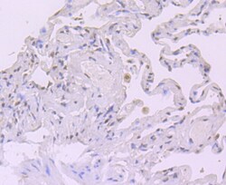
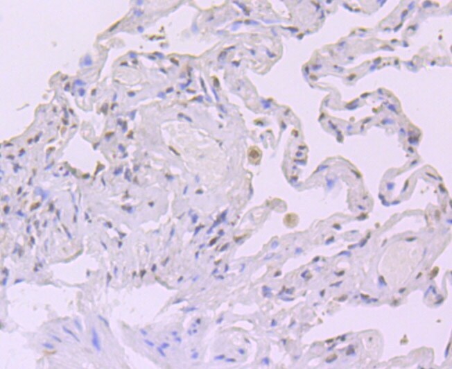
- Immunohistochemistry (Paraffin) analysis of CRM1 in paraffin-embedded human lung cancer tissue using CRM1 Recombinant Monoclonal Antibody (Product # MA5-41253). Counter stained with hematoxylin.
- Submitted by
- Invitrogen Antibodies (provider)
- Main image
- Experimental details
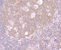
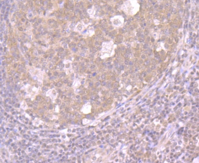
- Immunohistochemistry (Paraffin) analysis of CRM1 in paraffin-embedded human tonsil tissue using CRM1 Recombinant Monoclonal Antibody (Product # MA5-41253). Counter stained with hematoxylin.
- Submitted by
- Invitrogen Antibodies (provider)
- Main image
- Experimental details
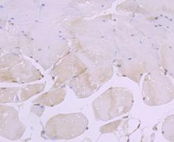
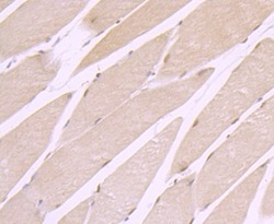
- Immunohistochemistry (Paraffin) analysis of CRM1 in paraffin-embedded mouse skeletal muscle tissue using CRM1 Recombinant Monoclonal Antibody (Product # MA5-41253). Counter stained with hematoxylin.
- Submitted by
- Invitrogen Antibodies (provider)
- Main image
- Experimental details
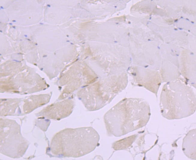
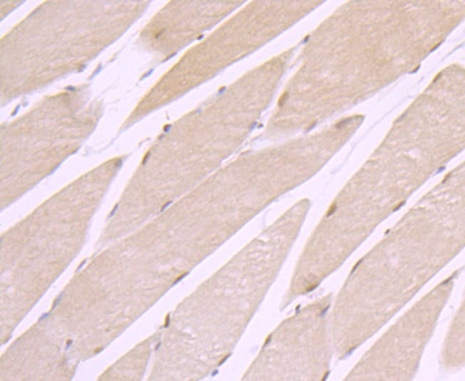
- Immunohistochemistry (Paraffin) analysis of CRM1 in paraffin-embedded rat skeletal muscle tissue using CRM1 Recombinant Monoclonal Antibody (Product # MA5-41253). Counter stained with hematoxylin.
Supportive validation
- Submitted by
- Invitrogen Antibodies (provider)
- Main image
- Experimental details
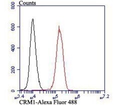
- Flow Cytometry analysis of CRM1 in K562 cells. The cells were fixed, permeabilized and stained with CRM1 Recombinant Monoclonal Antibody (Product # MA5-41253) at a dilution of 1:100 (red). Alexa Fluor 488-conjugated goat anti rabbit IgG was used as the secondary antibody. Unlabelled control (cells without incubation with primary antibody; black).
- Submitted by
- Invitrogen Antibodies (provider)
- Main image

- Experimental details
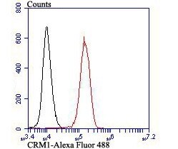
- Flow Cytometry analysis of CRM1 in K562 cells. The cells were fixed, permeabilized and stained with CRM1 Recombinant Monoclonal Antibody (Product # MA5-41253) at a dilution of 1:100 (red). Alexa Fluor 488-conjugated goat anti rabbit IgG was used as the secondary antibody. Unlabelled control (cells without incubation with primary antibody; black).
