Explore
 Validate
 Learn
Learn Western blot
Antibody data
- Antibody Data
- Antigen structure
- References [0]
- Comments [0]
- Validations
- Western blot [1]
- Immunocytochemistry [1]
- Immunohistochemistry [2]
- Flow cytometry [1]
Submit
Validation data
Reference
Comment
Report error
- Product number
- Ab131343 - Provider product page

- Provider
- Aladdin Scientific
- Product name
- TIMP2 Mouse mAb
- Antibody type
- Monoclonal
- Description
- Mouse anti Human TIMP2 Antibody, Monoclonal (1554CT494.262.47),Êcould be used for WB, IHC, ICC, IF, Flow and so on.ApplicationWB: 1/2000IHC: 1/200IF: 1/25Flow: 1/25Protein FunctionComplexes with metalloproteinases (such as collagenases) and irreversibly inactivates them by binding to their catalytic zinc cofactor. Known to act on MMP-1, MMP-2, MMP-3, MMP-7, MMP-8, MMP-9, MMP-10, MMP-13, MMP-14, MMP-15, MMP-16 and MMP-19.
- Reactivity
- Human, Mouse, Rat
- Host
- Mouse
- Conjugate
- Unconjugated
- Antigen sequence
AA 1-220- Antibody clone number
- 1554CT494.262.47
- Vial size
- 100_l,10_l,1ml,50_l
- Concentration
- 0,5 mg/ml
- Storage
- Store at 4_ short term (1-2 weeks). Store at -20_ long term (24 months). Upon delivery aliquot. Avoid freeze/thaw cycle.
No comments: Submit comment
Supportive validation
- Submitted by
- Aladdin Scientific (provider)
- Main image
- Experimental details
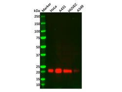
- TIMP2 Mouse mAb (Ab131343) - Western Blot All lanes: TIMP2 Mouse mAb (Ab131343) at 1/2000 dilution Samples: Lysates at 20 µg per lane Secondary: Goat Anti-Mouse IgG H&L (HRP) (Ab138040) at 1/20000 dilution Predicted band size: 24 kDa Observed band size: 21 kDa Exposure time: 120.0 s

Supportive validation
- Submitted by
- Aladdin Scientific (provider)
- Main image
- Experimental details
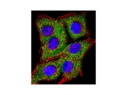
- TIMP2 Mouse mAb (Ab131343) - IF/ICC Immunofluorescent analysis of 4% paraformaldehyde-fixed, 0.1% Triton X-100 permeabilized A549 (human lung adenocarcinoma epithelial cell line) cells labeling TIMP2 with TIMP2 Mouse mAb (Ab131343) at 1/25 dilution, followed by Dylight® 488-conjugated goat anti-mouse IgG secondary antibody at 1/200 dilution (green). Immunofluorescence image showing cytoplasm staining on A549 cell line. Cytoplasmic actin is detected with Dylight® 554 Phalloidin at 1/100 dilution (red). The nuclear counter stain is DAPI (blue).
Supportive validation
- Submitted by
- Aladdin Scientific (provider)
- Main image
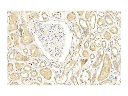
- Experimental details
- TIMP2 Mouse mAb (Ab131343) - IHC Immunohistochemical analysis of paraffin-embedded Human kidney section using TIMP2 Mouse mAb (Ab131343). TIMP2 Mouse mAb (Ab131343) was diluted at 1/200 dilution. A undiluted biotinylated goat polyvalent antibody was used as the secondary, followed by DAB staining.
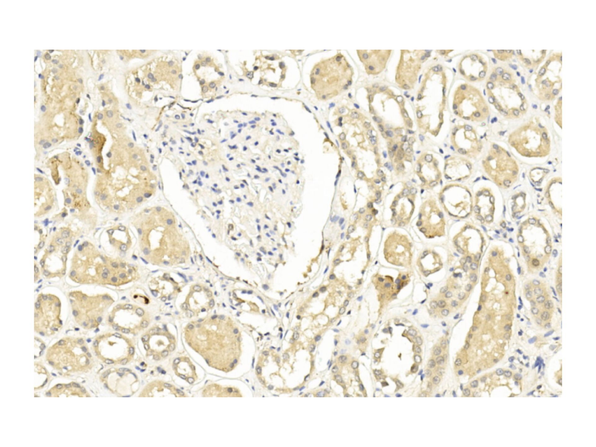
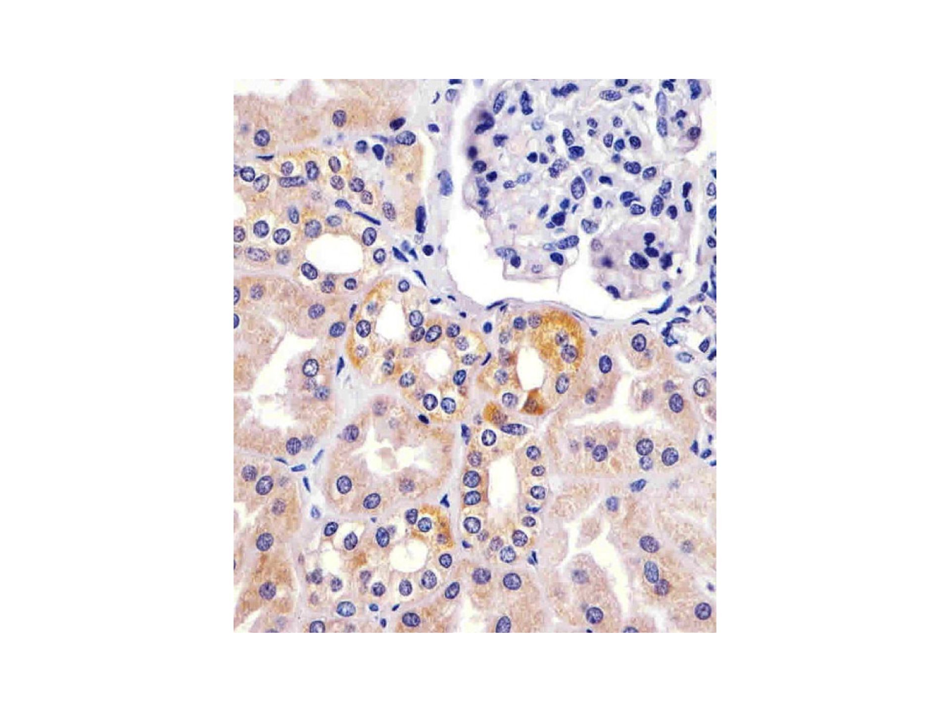
- Submitted by
- Aladdin Scientific (provider)
- Main image

- Experimental details
- TIMP2 Mouse mAb (Ab131343) - IHC TIMP2 Mouse mAb (Ab131343) staining TIMP2 in human kidney sections by Immunohistochemistry. Tissue was fixed with formaldehyde and blocked with 3% BSA for 0.5 hour at room temperature; antigen retrieval was by heat mediation with a citrate buffer (pH 6.0). Samples were incubated with TIMP2 Mouse mAb (Ab131343) (1/25) for 1 hours at 37°C. A undiluted biotinylated goat polyvalent antibody was used as the secondary antibody.
Supportive validation
- Submitted by
- Aladdin Scientific (provider)
- Main image
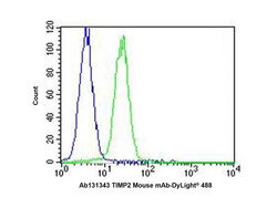
- Experimental details
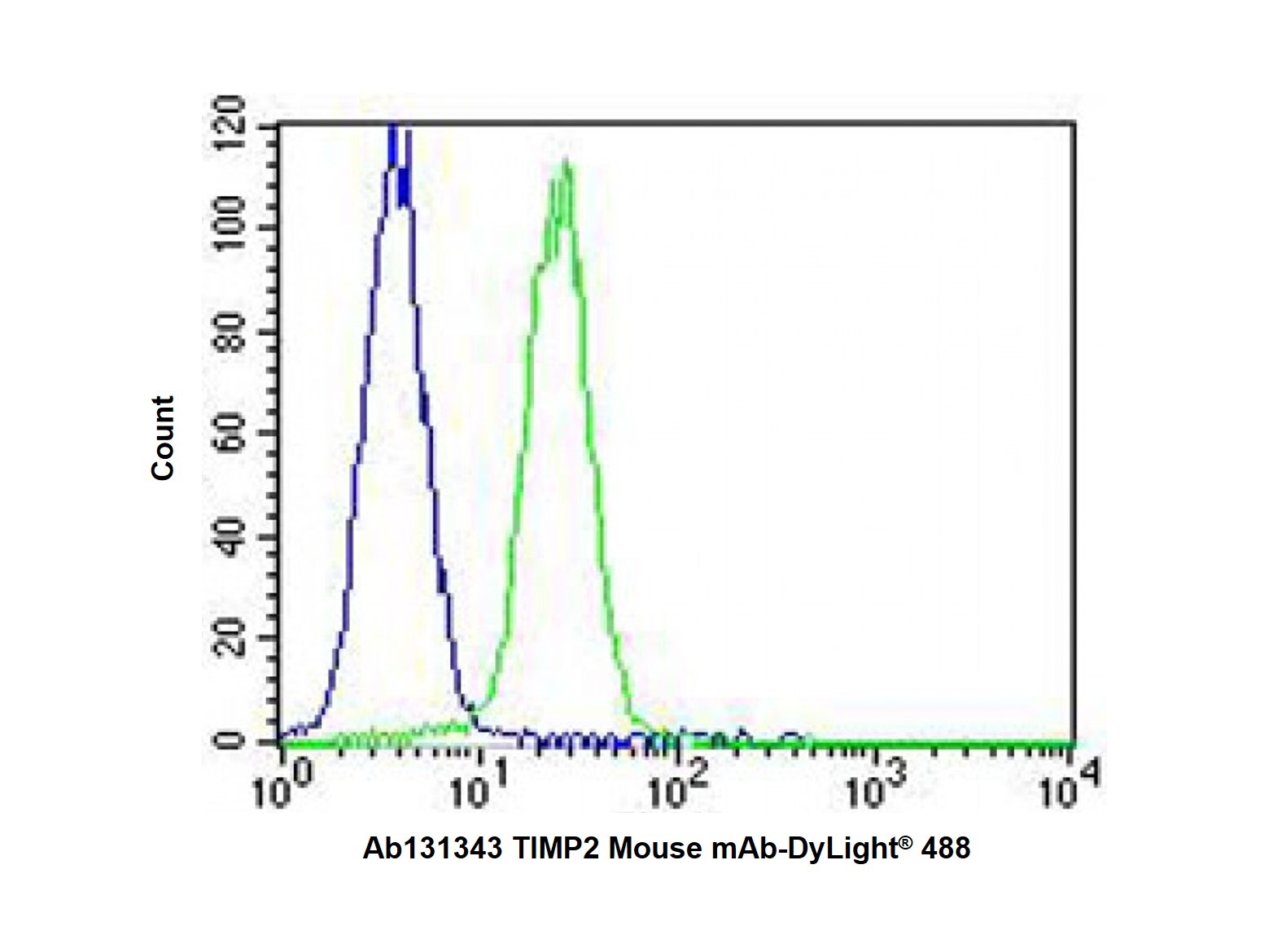
- TIMP2 Mouse mAb (Ab131343) - Flow Cytometry Overlay histogram showing K562 cells stained with TIMP2 Mouse mAb (Ab131343) (green line). The cells were fixed with 2% paraformaldehyde (10 min) and then permeabilized with 90% methanol for 10 min. The cells were then icubated in 2% bovine serum albumin to block non-specific protein-protein interactions followed by the TIMP2 Mouse mAb (Ab131343) (1/25 dilution) for 60 min at 37ºC. The secondary antibody used was Goat-Anti-Mouse IgG, DyLight® 488 Conjugated Highly Cross-Adsorbed at 1/400 dilution for 40 min at 37ºC. Isotype control antibody (blue line) was mouse IgG1 (1μg/1x10^6 cells) used under the same conditions. Acquisition of >10000 events was performed.