 Explore
 Validate
 Learn
Learn Western blot
 Immunocytochemistry
Antibody data
- Antibody Data
- Antigen structure
- References [1]
- Comments [0]
- Validations
- Immunocytochemistry [1]
- Flow cytometry [4]
Submit
Validation data
Reference
Comment
Report error
- Product number
- MA1-81548 - Provider product page

- Provider
- Invitrogen Antibodies
- Product name
- IGF1R alpha Monoclonal Antibody (1H7)
- Antibody type
- Monoclonal
- Antigen
- Purifed from natural sources
- Description
- This product requires protein digestion pre-treatment of paraffin sections using trypsin or pronase. A suggested positive control for immunohistochemical applications is pancreas or placenta. For FACS analysis, use 10 µL of the suggested working dilution to label 1x10^6 cells in 100 µL. Mouse anti Human CD221 antibody, clone 1H7 recognizes human CD221, a approximately 155 kDa receptor tyrosine kinase, also known as Insulin-like growth factor I receptor (IGF-I Receptor).
- Reactivity
- Human
- Host
- Mouse
- Isotype
- IgG
- Antibody clone number
- 1H7
- Vial size
- 200 µg
- Concentration
- 1 mg/mL
- Storage
- Store at 4°C short term. For long term storage, store at -20°C, avoiding freeze/thaw cycles.
Submitted references Differential activation of a C/EBP beta isoform by a novel redox switch may confer the lipopolysaccharide-inducible expression of interleukin-6 gene.
Su WC, Chou HY, Chang CJ, Lee YM, Chen WH, Huang KH, Lee MY, Lee SC
The Journal of biological chemistry 2003 Dec 19;278(51):51150-8
The Journal of biological chemistry 2003 Dec 19;278(51):51150-8
No comments: Submit comment
Supportive validation
- Submitted by
- Invitrogen Antibodies (provider)
- Main image
- Experimental details
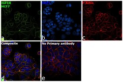
- Immunofluorescence analysis of Insulin-like growth factor 1 receptor was performed using 70% confluent log phase MCF7 cells. The cells were fixed with 4% paraformaldehyde for 10 minutes, permeabilized with 0.1% Triton™ X-100 for 15 minutes, and blocked with 2% BSA for 45 minutes at room temperature. The cells were labeled with IGF1R alpha Monoclonal Antibody (1H7) (Product # MA1-81548) at 1:100 in 0.1% BSA, incubated at 4 degree celsius overnight and then labeled with Goat anti-Mouse IgG (H+L) Highly Cross-Adsorbed Secondary Antibody, Alexa Fluor Plus 488 (Product # A32723), (1:2000), for 45 minutes at room temperature (Panel a: Green). Nuclei (Panel b:Blue) were stained with ProLong™ Diamond Antifade Mountant with DAPI (Product # P36962). F-actin (Panel c: Red) was stained with Rhodamine Phalloidin (Product # R415, 1:300). Panel d represents the merged image showing Membrane localization. Panel e represents control cells with no primary antibody to assess background. The images were captured at 60X magnification.

Supportive validation
- Submitted by
- Invitrogen Antibodies (provider)
- Main image
- Experimental details
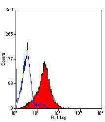
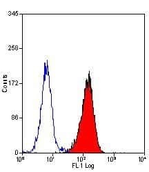
- Flow cytometric analysis of human peripheral blood granulocytes using a CD221/IGF1 Receptor monoclonal antibody (Product # MA1-81548)

- Submitted by
- Invitrogen Antibodies (provider)
- Main image
- Experimental details
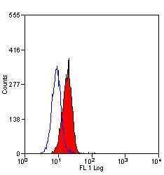
- Flow cytometric analysis of human peripheral blood granulocytes using a CD221/IGF1 Receptor monoclonal antibody (Product # MA1-81548)

- Submitted by
- Invitrogen Antibodies (provider)
- Main image
- Experimental details
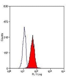
- Flow cytometric analysis of human peripheral blood granulocytes using a CD221/IGF1 Receptor monoclonal antibody (Product # MA1-81548)

- Submitted by
- Invitrogen Antibodies (provider)
- Main image
- Experimental details
- Flow cytometric analysis of human peripheral blood granulocytes using a CD221/IGF1 Receptor monoclonal antibody (Product # MA1-81548)
