 Explore
 Validate
 Learn
Learn Western blot
 ELISA
ELISAAntibody data
- Antibody Data
- Antigen structure
- References [4]
- Comments [0]
- Validations
- Western blot [3]
- Other assay [7]
Submit
Validation data
Reference
Comment
Report error
- Product number
- 39-6700 - Provider product page

- Provider
- Invitrogen Antibodies
- Product name
- IGF1R beta Monoclonal Antibody (ZI001)
- Antibody type
- Monoclonal
- Antigen
- Other
- Reactivity
- Human
- Host
- Mouse
- Isotype
- IgG
- Antibody clone number
- ZI001
- Vial size
- 100 µg
- Concentration
- 0.5 mg/mL
- Storage
- -20°C
Submitted references The transcription factors Ik-1 and MZF1 downregulate IGF-IR expression in NPM-ALK⁺ T-cell lymphoma.
The ALK inhibitor ASP3026 eradicates NPM-ALK⁺ T-cell anaplastic large-cell lymphoma in vitro and in a systemic xenograft lymphoma model.
MicroRNA 96 is a post-transcriptional suppressor of anaplastic lymphoma kinase expression.
Inhibition of IGF-IR tyrosine kinase induces apoptosis and cell cycle arrest in imatinib-resistant chronic myeloid leukaemia cells.
Vishwamitra D, Curry CV, Alkan S, Song YH, Gallick GE, Kaseb AO, Shi P, Amin HM
Molecular cancer 2015 Feb 25;14:53
Molecular cancer 2015 Feb 25;14:53
The ALK inhibitor ASP3026 eradicates NPM-ALK⁺ T-cell anaplastic large-cell lymphoma in vitro and in a systemic xenograft lymphoma model.
George SK, Vishwamitra D, Manshouri R, Shi P, Amin HM
Oncotarget 2014 Jul 30;5(14):5750-63
Oncotarget 2014 Jul 30;5(14):5750-63
MicroRNA 96 is a post-transcriptional suppressor of anaplastic lymphoma kinase expression.
Vishwamitra D, Li Y, Wilson D, Manshouri R, Curry CV, Shi B, Tang XM, Sheehan AM, Wistuba II, Shi P, Amin HM
The American journal of pathology 2012 May;180(5):1772-80
The American journal of pathology 2012 May;180(5):1772-80
Inhibition of IGF-IR tyrosine kinase induces apoptosis and cell cycle arrest in imatinib-resistant chronic myeloid leukaemia cells.
Shi P, Chandra J, Sun X, Gergely M, Cortes JE, Garcia-Manero G, Arlinghaus RB, Lai R, Amin HM
Journal of cellular and molecular medicine 2010 Jun;14(6B):1777-92
Journal of cellular and molecular medicine 2010 Jun;14(6B):1777-92
No comments: Submit comment
Supportive validation
- Submitted by
- Invitrogen Antibodies (provider)
- Main image
- Experimental details
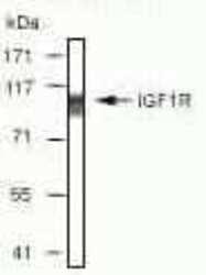
- Western blot analysis of MCF7 cell extract using Zymed Ms anti-IGF1Rbeta (Product # 39-6700).

- Submitted by
- Invitrogen Antibodies (provider)
- Main image

- Experimental details
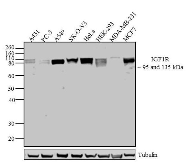
- Western blot analysis of IGF-IR/IGF1 Receptor was performed by loading 30 µg of A431 (lane1), PC-3 (lane2), A549 (lane3), SK-O-V3 (lane4), HeLa (lane5), HEK-293 (lane6), MDA-MB-231 (lane7) and MCF7 (lane8) cell lysate using NuPAGE® Novex® 10% Bis-Tris gel (Product # NP0301BOX), XCell SureLock™ Electrophoresis System (Product # EI0002), Novex® Sharp Pre-Stained Protein Standard (LC5800). Proteins were transferred to a PVDF membrane and blocked with 5% skim milk for 1 hour at room temperature. IGF-IR/IGF1 Receptor was detected at 95 and 135 kDa using IGF-IR/IGF1 Receptor Mouse Monoclonal Antibody (Product # 39-6700) at 1:500 dilution in 2.5% skim milk at 4°C overnight on a rocking platform. Goat Anti-Mouse - HRP Secondary Antibody (Product # 62-6520) at 1:4000 dilution was used and chemiluminescent detection was performed using Pierce™ ECL Western Blotting Substrate (Product # 32106). Additional bands at 135 and 95 kDa represent the alpha and beta chains of a fully mature cell membrane-bound IGF1R.

- Submitted by
- Invitrogen Antibodies (provider)
- Main image

- Experimental details
- Western blot analysis of IGF-IR/IGF1 Receptor was performed by loading 30 µg of A431 (lane1), PC-3 (lane2), A549 (lane3), SK-O-V3 (lane4), HeLa (lane5), HEK-293 (lane6), MDA-MB-231 (lane7) and MCF7 (lane8) cell lysate using NuPAGE® Novex® 10% Bis-Tris gel (Product # NP0301BOX), XCell SureLock™ Electrophoresis System (Product # EI0002), Novex® Sharp Pre-Stained Protein Standard (LC5800). Proteins were transferred to a PVDF membrane and blocked with 5% skim milk for 1 hour at room temperature. IGF-IR/IGF1 Receptor was detected at 95 and 135 kDa using IGF-IR/IGF1 Receptor Mouse Monoclonal Antibody (Product # 39-6700) at 1:500 dilution in 2.5% skim milk at 4°C overnight on a rocking platform. Goat Anti-Mouse - HRP Secondary Antibody (Product # 62-6520) at 1:4000 dilution was used and chemiluminescent detection was performed using Pierce™ ECL Western Blotting Substrate (Product # 32106). Additional bands at 135 and 95 kDa represent the alpha and beta chains of a fully mature cell membrane-bound IGF1R.
Supportive validation
- Submitted by
- Invitrogen Antibodies (provider)
- Main image
- Experimental details
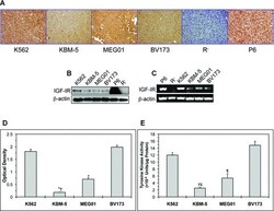
- Fig 1 Expression of IGF-IR in CML cell lines. (A) Immunohistochemical staining shows strong and universal expression of IGF-IR in four CML cell lines including K562, KBM-5, MEG01 and BV173. (B) Western blotting confirms the expression of IGF-IR protein in all of the CML cell lines. The K562 cell line demonstrates a higher level of IGF-IR protein compared with the other three cell lines. beta-Actin confirms equal loading. (C) RT-PCR shows the expression of IGF-IR mRNA in CML cell lines. Similar to IGF-IR protein levels, K562 cells appear to express the highest level of IGF-IR mRNA. beta-Actin supports equal loading. In all of these experiments, R - and P6 cells served as negative and positive controls for the expression of IGF-IR, respectively. (D) The KBM-5 and MEG01 cell lines demonstrate lower levels of pIGF-IR in comparison with K562 and BV173. The results represent the means +- S.D. of three experiments. *: P < 0.0001 compared with K562 and BV173 and +: P < 0.0001 compared with MEG01. (E) The KBM-5 and MEG01 cell lines possess the lowest levels of IGF-IR tyrosine kinase activity. Depicted results represent the means +- S.D. of three experiments. *: P < 0.05 compared with BV173, +: P < 0.0001 compared with K562 and BV173, ++: P < 0.05 compared with MEG01 and P: P < 0.001 compared with K562 and BV173.

- Submitted by
- Invitrogen Antibodies (provider)
- Main image

- Experimental details
- Fig 2 Expression of IGF-IR in primary human CML specimens. (A) A representative bone marrow specimen from a patient with CML in BP demonstrates strong expression of IGF-IR protein compared with bone marrow specimen from a CML patient in CP that is negative for IGF-IR. (B) The markedly increased expression of IGF-IR in the majority of BP CML patients (four out of five patients) compared with patients in CP (four patients) or AP (five patients) is also demonstrated by real-time quantitative PCR analysis of primary neoplastic cells.

- Submitted by
- Invitrogen Antibodies (provider)
- Main image

- Experimental details
- Fig 5 Inhibition of IGF-IR decreases the viability of imatinib-resistant p210 BCR-ABL-expressing cells. (A) Western blotting shows the expression of IGF-IR, pIGF-IR, WT p210 BCR-ABL, BCR-ABL E255K or BCR-ABL T315I in BaF3 cells. BaF3 cells transfected with empty vector lack the expression of BCR-ABL. beta-Actin confirms equal loading of the proteins. (B) Only BaF3 cells transfected with WT p210 BCR-ABL demonstrate time-dependent decrease in cell viability after treatment with imatinib (5.0 muM). In contrast, BaF3 cells transfected with BCR-ABL mutants or empty vector are completely resistant to imatinib treatment. *: P < 0.001 compared with control untreated cells and +: P < 0.001 compared with imatinib (5.0 muM; 24 hrs). (C) Targeting IGF-IR by PPP induces concentration- and time-dependent decrease in the viability of BaF3 cells transfected with WT BCR-ABL or the BCR-ABL E255K and BCR-ABL T315I mutants that are known to be resistant to imatinib. The results at 24 hrs are shown in the left panel and those at 48 hrs in the right panel. The results are means +- S.D. of three consistent experiments. *: P < 0.05 and +: P < 0.001 versus control untreated cells. (D) Treatment of KBM-5 or BV173 cell lines with PPP alone is significantly more effective in decreasing the viability of these cells than treatment with imatinib alone. The effect on cell viability of all CML cell lines is significantly enhanced when imatinib was combined with PPP. Notably, the MEG01 cell line demonstrates

- Submitted by
- Invitrogen Antibodies (provider)
- Main image

- Experimental details
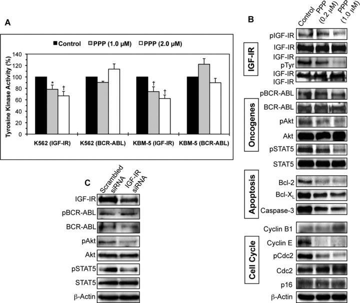
- Fig 6 Effects of inhibition of IGF-IR on IGF-IR, BCR-ABL and downstream target proteins in CML cell lines. (A) PPP induces concentration-dependent decrease in IGF-IR tyrosine kinase activity in K562 and KBM-5 cell lines. In contrast, PPP fails to cause similar effect in BCR-ABL tyrosine kinase activity. The results represent the means +- S.D. of three consistent experiments. *: P < 0.01 and +: P < 0.001 compared with control untreated cells. (B) Western blotting and co-immunoprecipitation studies confirm that PPP decreases the tyrosine phosphorylation of IGF-IR in a concentration-dependent fashion (results shown are representative and were obtained from the KBM-5 cell line). The basal levels of IGF-IR did not change after treatment with PPP. The phosphorylation level of BCR-ABL remains unchanged after treatment with PPP. The decrease in pIGF-IR is associated with down-regulation of pAkt and pSTAT5, two oncogenic proteins in CML. Changes are not seen in Akt and STAT5. Also, PPP induces changes consistent with apoptotic cell death including down-regulation of Bcl-2, Bcl-X L and caspase-3. Moreover, treatment with PPP induces up-regulation of cyclin B1 and down-regulation of cyclin E and pCdc2, whereas the levels of Cdc2 and p16 remain unchanged. Overall, the changes in the cell cycle regulatory proteins are consistent with G2/M-phase cell cycle arrest. beta-Actin shows equal loading of the proteins. (C) IGF-IR siRNA decreases IGF-IR levels in the KBM-5 cell line and this decrea
- Submitted by
- Invitrogen Antibodies (provider)
- Main image
- Experimental details
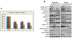
- Figure 2 ASP3026 reduces the tyrosine kinase activity of NPM-ALK, downregulates the phosphorylation of NPM-ALK and target proteins, and induces biochemical effects consistent with apoptosis (A) ASP3026 induced a marked decrease in NPM-ALK tyrosine kinase activity in the lymphoma cells (*: P < 0.01, **: P < 0.001, ***: P < 0.0001 compared with control cells treated with vehicle). Results shown are means +- SE of 3 experiments. (B) ASP3026 decreased the phosphorylation of NPM-ALK at 2 different tyrosine residues: Y646 and Y664. Also, treatment with ASP3026 was associated with downregulation of the phosphorylation of the NPM-ALK target proteins IGF-IR, STAT3, AKT, and JNK. Changes in the basal levels of these proteins were not detected. The occurrence of apoptosis was biochemically supported by the increase in cleaved caspase-3 and cleaved PARP and the simultaneous decrease in non-cleaved caspase-3 and non-cleared PARP levels. beta-Actin indicates equal loading of the proteins.

- Submitted by
- Invitrogen Antibodies (provider)
- Main image

- Experimental details
- Figure 3 Ik-1 and MZF1 decrease IGF-IR mRNA and protein levels and the phosphorylation of downstream targets. (A) Western blotting demonstrates increased expression levels of Ik-1 and MZF1 proteins at 48 h after transfection into 3 NPM-ALK + T-cell lymphoma cell lines. beta-actin shows equal protein loading (-: transfection of EV; +: transfection of Ik-1 or MZF1). (B) Transfection of Ik-1 remarkably decreased IGF-IR mRNA levels in Karpas 299, SUP-M2, and SR-786 cell lines (*: < 0.0001 compared with EV). (C) Similarly, transfection of MZF1 induced a significant decrease in IGF-IR mRNA levels (*: < 0.0001 compared with EV). The results depicted in (B) and (C) represent the means +- SE of 3 experiments. (D) Western blotting shows that transfection of Ik-1 and MZF1 into Karpas 299, SUP-M2, and SR-786 cell lines induced marked downregulation of IGF-IR protein, which was associated with decreased pIGF-IR levels. Moreover, the decrease in IGF-IR/pIGF-IR expression levels was associated with decreased phosphorylation of important IGF-IR targets including IRS-1, AKT, and NPM-ALK. Whereas basal levels of AKT remained unchanged, notable decrease in IRS-1 protein was observed. The 3 web-based transcription factor search algorithms showed that Ik-1 and MZF1 could potentially bind the IRS-1 gene promoter. In contrast, searching these algorithms did not support potential binding of Ik-1 or MZF1 and the NPM gene promoter, where the expression of NPM-ALK protein is regulated at the transcript

- Submitted by
- Invitrogen Antibodies (provider)
- Main image

- Experimental details
- Figure 6 NPM-ALK oncogenic protein does not affect the levels of expression of IGF-IR and IGF-I. (A) Western blotting shows that at 48 h, downregulation of NPM-ALK by ALK siRNA was not associated with decreased expression of IGF-IR, pro-IGF-I or IGF-I proteins in SUP-M2, SR-786, and DEL cell lines. beta-Actin shows equal protein loading. Analysis of IGF-IR levels after transfection of the cells with ALK siRNA was performed at extended time points (12, 24, 48, 72, and 96 h), and also in other cell lines including Karpas 299 and SU-DHL-1, with similar results (data not shown). (B) Downregulation of NPM-ALK in the 3 cell lines did not decrease the levels of IGF-IR mRNA. The example shown is at 48 h after transfection of the cells with ALK siRNA. The results are shown as means +- SE of 4 consistent experiments. In addition, analysis of IGF-IR mRNA was performed at other time points and cell lines as described in (A). Changes in IGF-IR mRNA levels were not detected at any time point (data not shown). (C) An ELISA assay showing that specific downregulation of NPM-ALK did not significantly decrease the levels of IGF-I secreted from the NPM-ALK + T-cell lymphoma cells. The results represent the means +- SE of 3 experiments.
