 Explore
 Validate
 Learn
Learn Western blot
 ELISA
ELISAAntibody data
- Antibody Data
- Antigen structure
- References [0]
- Comments [0]
- Validations
- Western blot [1]
- Immunohistochemistry [1]
Submit
Validation data
Reference
Comment
Report error
- Product number
- AP14935PU-N - Provider product page

- Provider
- Acris Antibodies GmbH
- Proper citation
- Acris Antibodies GmbH Cat#AP14935PU-N, RRID:AB_1770506
- Product name
- anti PIK3CG (N-term)
- Antibody type
- Polyclonal
- Antigen
- This antibody is generated from rabbits immunized with a KLH conjugated synthetic peptide selected from the N-terminal region of human PI3KCG.
- Reactivity
- Human, Mouse
- Host
- Rabbit
- Vial size
- 0.1 mg
- Concentration
- 0.25 mg/ml
No comments: Submit comment
Supportive validation
- Submitted by
- Acris Antibodies GmbH (provider)
- Main image
- Experimental details
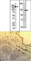
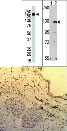
- (TOP LEFT) The anti-PI3KCG Pab is used in Western blot to detect PI3KCG in mouse skeletal muscle tissue lysate. (TOP RIGHT)Western blot analysis of PI3KCG (arrow) using rabbit polyclonal PI3KCG Antibody (N-term). 293 cell lysates (2 ug/lane) either nontransfected (Lane 1) or transiently transfected with the PI3KCG gene (Lane 2) (Origene Technologies). (BOTTOM)This data demonstrates the use of PI3KCG antibody for IHC. Frozen tissue from the mouse ear reacted with the PI3KCG antibody, which was detected by peroxidase-conjugated anti-rabbit secondary antibody and staining with Vesctain Elite ABC/DAB kit. Sections were counterstained with hematoxilin.
Supportive validation
- Submitted by
- Acris Antibodies GmbH (provider)
- Main image
- Experimental details
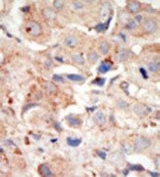
- Formalin-fixed and paraffin-embedded human cancer tissue reacted with the primary antibody, which was peroxidase-conjugated to the secondary antibody, followed by DAB staining.
