Explore
 Validate
 Learn
Learn Western blot
 Immunocytochemistry
Antibody data
- Antibody Data
- Antigen structure
- References [3]
- Comments [0]
- Validations
- Immunocytochemistry [1]
- Immunohistochemistry [1]
Submit
Validation data
Reference
Comment
Report error
- Product number
- HPA015257 - Provider product page

- Provider
- Atlas Antibodies
- Proper citation
- Atlas Antibodies Cat#HPA015257, RRID:AB_1856294
- Product name
- Anti-RIPK1
- Antibody type
- Polyclonal
- Description
- Polyclonal Antibody against Human RIPK1, Gene description: receptor (TNFRSF)-interacting serine-threonine kinase 1, Alternative Gene Names: RIP, Validated applications: IHC, WB, ICC, Uniprot ID: Q13546, Storage: Store at +4°C for short term storage. Long time storage is recommended at -20°C.
- Reactivity
- Human
- Host
- Rabbit
- Conjugate
- Unconjugated
- Isotype
- IgG
- Vial size
- 100 µl
- Concentration
- 0.2 mg/ml
- Storage
- Store at +4°C for short term storage. Long time storage is recommended at -20°C.
- Handling
- The antibody solution should be gently mixed before use.
Submitted references RIPK1 and RIPK3 are positive prognosticators for cervical cancer patients and C2 ceramide can inhibit tumor cell proliferation in vitro
Acetylcholine and necroptosis are players in follicular development in primates
RIP1 expression is necessary for CD30-mediated cell death induction in anaplastic large-cell lymphoma cells
Vogelsang T, Kast V, Bagnjuk K, Eubler K, Jeevanandan S, Schmoeckel E, Trebo A, Topalov N, Mahner S, Mayr D, Mayerhofer A, Jeschke U, Vattai A
Frontiers in Oncology 2023;13
Frontiers in Oncology 2023;13
Acetylcholine and necroptosis are players in follicular development in primates
Du Y, Bagnjuk K, Lawson M, Xu J, Mayerhofer A
Scientific Reports 2018;8(1)
Scientific Reports 2018;8(1)
RIP1 expression is necessary for CD30-mediated cell death induction in anaplastic large-cell lymphoma cells
Hirsch B, von der Wall E, Hummel M, Dürkop H
Laboratory Investigation 2013;93(6):677-689
Laboratory Investigation 2013;93(6):677-689
No comments: Submit comment
Supportive validation
- Submitted by
- Atlas Antibodies (provider)
- Main image

- Experimental details
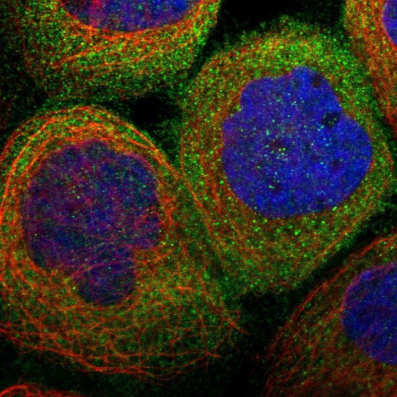
- Immunofluorescent staining of human cell line A-431 shows localization to cytosol.
- Sample type
- Human
Supportive validation
- Submitted by
- Atlas Antibodies (provider)
- Enhanced method
- Orthogonal validation
- Main image
- Experimental details
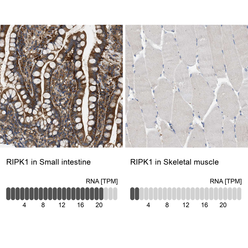
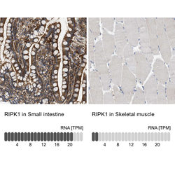
- Immunohistochemistry analysis in human small intestine and skeletal muscle tissues using HPA015257 antibody. Corresponding RIPK1 RNA-seq data are presented for the same tissues.
- Sample type
- Human
- Protocol
- Protocol