 Explore
 Validate
 Learn
Learn Western blot
Antibody data
- Antibody Data
- Antigen structure
- References [1]
- Comments [0]
- Validations
- Western blot [1]
- ELISA [1]
- Immunohistochemistry [1]
- Flow cytometry [2]
- Other assay [1]
Submit
Validation data
Reference
Comment
Report error
- Product number
- 701080 - Provider product page

- Provider
- Invitrogen Antibodies
- Product name
- IL-2 Recombinant Rabbit Monoclonal Antibody (2H20L7)
- Antibody type
- Monoclonal
- Antigen
- Recombinant full-length protein
- Description
- Intact IgG appears on a non-reducing gel as ~150 kDa band and upon reduction generating a ~25 kDa light chain band and a ~50 kDa heavy chain. 701080 was successfully to detect IL-2 in Flow and ELISA application. Recombinant rabbit monoclonal antibodies are produced using in vitro expression systems. The expression systems are developed by cloning in the specific antibody DNA sequences from immunoreactive rabbits. Then, individual clones are screened to select the best candidates for production. The advantages of using recombinant rabbit monoclonal antibodies include: better specificity and sensitivity, lot-to-lot consistency, animal origin-free formulations, and broader immunoreactivity to diverse targets due to larger rabbit immune repertoire.
- Reactivity
- Human
- Host
- Rabbit
- Isotype
- IgG
- Antibody clone number
- 2H20L7
- Vial size
- 100 μg
- Concentration
- 0.5 mg/mL
- Storage
- Store at 4°C short term. For long term storage, store at -20°C, avoiding freeze/thaw cycles.
Submitted references RIPK3 and Caspase-1/11 Are Necessary for Optimal Antigen-Specific CD8 T Cell Response Elicited by Genetically Modified Listeria monocytogenes.
Rana A, de Almeida FC, Paico Montero HA, Gonzales Carazas MM, Bortoluci KR, Sad S, Amarante-Mendes GP
Frontiers in immunology 2020;11:536
Frontiers in immunology 2020;11:536
No comments: Submit comment
Supportive validation
- Submitted by
- Invitrogen Antibodies (provider)
- Main image
- Experimental details
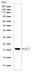
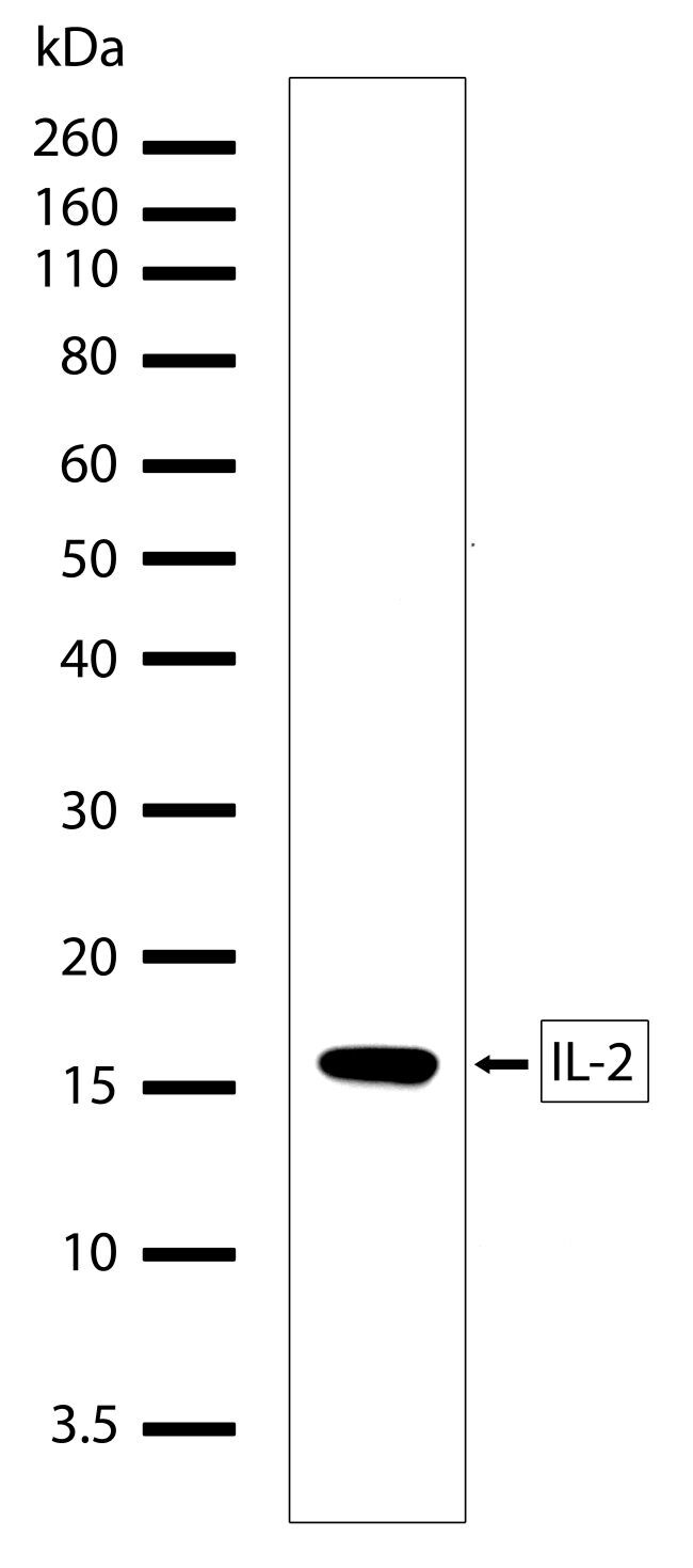
- Western blot analysis of recombinant human IL-2 protein using an IL-2 recombinant rabbit monoclonal antibody (Product # 701080).
Supportive validation
- Submitted by
- Invitrogen Antibodies (provider)
- Main image
- Experimental details
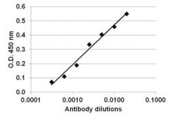
- Direct ELISA analysis of IL-2 was performed by coating wells of a 96-well plate with 100 µL per well of IL-2 (Product # RIL2I) diluted in carbonate/bicarbonate buffer (Product # 28382) at a concentration of 2 µg/mL overnight at 4C. Wells of the plate were washed, blocked with starting blocking buffer (Product # 37538), and incubated with 100 µL per well of a rabbit anti-IL-2 monoclonal antibody (Product # 701080) at a serially dilution of 1:50, 1:100, 1:200, 1:400, 1:800, 1:1600 and 1:3200 for 90 minutes at 37C. The plate was washed, then incubated with 100 µL per well of an HRP-conjugated goat anti-rabbit IgG secondary antibody (Product # 65-6120) at a dilution of 1:5000 for 90 minutes at 37C. Detection was performed using 1-Step Ultra TMB substrate (Product # 34028) for 5-10 minutes at room temperature in the dark. The reaction was stopped with Stop solution (Product # N600), and absorbances were read on a spectrophotometer at 450-550 nm.

Supportive validation
- Submitted by
- Invitrogen Antibodies (provider)
- Main image
- Experimental details
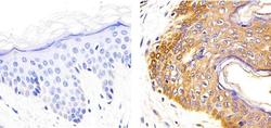
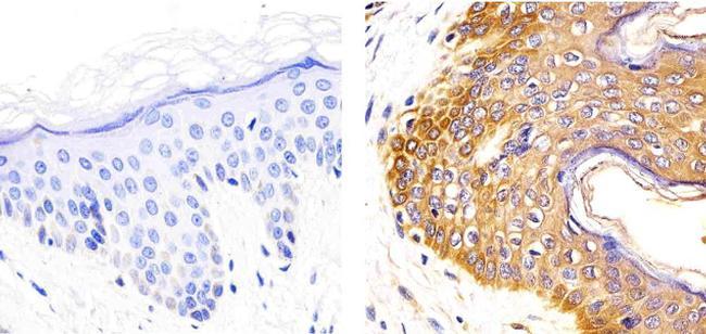
- Immunohistochemistry analysis of IL-2 showing staining in the cytoplasm of paraffin-embedded human skin tissue (right) compared to a negative control without primary antibody (left). To expose target proteins, antigen retrieval was performed using 10mM sodium citrate (pH 6.0), microwaved for 8-15 min. Following antigen retrieval, tissues were blocked in 3% H2O2-methanol for 15 min at room temperature, washed with ddH2O and PBS, and then probed with a IL-2 Recombinant Rabbit Monoclonal Antibody (Product # 701080) diluted in 3% BSA-PBS at a dilution of 1:20 overnight at 4°C in a humidified chamber. Tissues were washed extensively in PBST and detection was performed using an HRP-conjugated secondary antibody followed by colorimetric detection using a DAB kit. Tissues were counterstained with hematoxylin and dehydrated with ethanol and xylene to prep for mounting.
Supportive validation
- Submitted by
- Invitrogen Antibodies (provider)
- Main image

- Experimental details
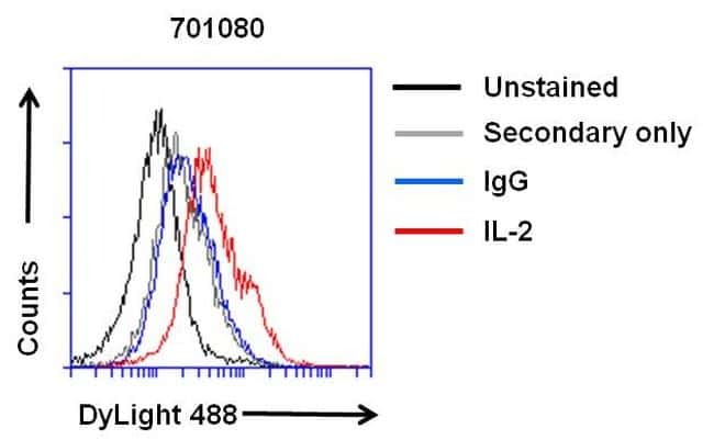
- Flow cytometry analysis of IL-2 on PHA-M treated Jurkat cells. Cells were fixed with 4% formaldehyde for 30 min on ince and permeabilized with IC permeabilization buffer (Product # PB001). After incubation with blocking buffer (Product # 37525) for 30 min on ice, cells were then stained with anti-IL-2 rabbit monoclonal antibody (Product # 701080) or IgG control at 1:100 dilution with IC permeabilization buffer for 30 min on ice. After washing with ice-cold IC permeabilization buffer for 3 times, the cells were stained with DyLight 488 goat anti-rabbit secondary antibody (Product # 35552) for 30 min on ice. A representative 10,000 cells were acquired for each sample.
- Submitted by
- Invitrogen Antibodies (provider)
- Main image

- Experimental details
- Flow cytometry analysis of IL-2 on PHA-M treated Jurkat cells. Cells were fixed with 4% formaldehyde for 30 min on ince and permeabilized with IC permeabilization buffer (Product # PB001). After incubation with blocking buffer (Product # 37525) for 30 min on ice, cells were then stained with anti-IL-2 rabbit monoclonal antibody (Product # 701080) or IgG control at 1:100 dilution with IC permeabilization buffer for 30 min on ice. After washing with ice-cold IC permeabilization buffer for 3 times, the cells were stained with DyLight 488 goat anti-rabbit secondary antibody (Product # 35552) for 30 min on ice. A representative 10,000 cells were acquired for each sample.

Supportive validation
- Submitted by
- Invitrogen Antibodies (provider)
- Main image

- Experimental details
- Direct ELISA analysis of IL-2 was performed by coating wells of a 96-well plate with 100 µL per well of IL-2 (Product # RIL2I) diluted in carbonate/bicarbonate buffer (Product # 28382) at a concentration of 2 µg/mL overnight at 4C. Wells of the plate were washed, blocked with starting blocking buffer (Product # 37538), and incubated with 100 µL per well of a rabbit anti-IL-2 monoclonal antibody (Product # 701080) at a serially dilution of 1:50, 1:100, 1:200, 1:400, 1:800, 1:1600 and 1:3200 for 90 minutes at 37C. The plate was washed, then incubated with 100 µL per well of an HRP-conjugated goat anti-rabbit IgG secondary antibody (Product # 65-6120) at a dilution of 1:5000 for 90 minutes at 37C. Detection was performed using 1-Step Ultra TMB substrate (Product # 34028) for 5-10 minutes at room temperature in the dark. The reaction was stopped with Stop solution (Product # N600), and absorbances were read on a spectrophotometer at 450-550 nm.
