Explore
 Validate
 Learn
Learn Western blot
 ELISA
ELISAAntibody data
- Antibody Data
- Antigen structure
- References [5]
- Comments [0]
- Validations
- Western blot [1]
- Immunocytochemistry [1]
- Immunohistochemistry [1]
- Blocking/Neutralizing [1]
Submit
Validation data
Reference
Comment
Report error
- Product number
- AF-502-NA - Provider product page

- Provider
- R&D Systems
- Product name
- Human/Rat IL-2 Antibody
- Antibody type
- Polyclonal
- Description
- Antigen Affinity-purified. Detects rat IL-2 in ELISAs. Detects human and rat IL-2 in Western blots. In sandwich immunoassays, less than 0.2% cross-reactivity with recombinant mouse IL-2 is observed.
- Reactivity
- Human, Rat
- Host
- Goat
- Conjugate
- Unconjugated
- Antigen sequence
P17108- Isotype
- IgG
- Vial size
- 100 ug
- Concentration
- LYOPH
- Storage
- Use a manual defrost freezer and avoid repeated freeze-thaw cycles. 12 months from date of receipt, -20 to -70 °C as supplied. 1 month, 2 to 8 °C under sterile conditions after reconstitution. 6 months, -20 to -70 °C under sterile conditions after reconstitution.
Submitted references A Crucial Role of CXCL14 for Promoting Regulatory T Cells Activation in Stroke.
Antidiabetic Effect of Interleukin-1β Antibody Therapy Through β-Cell Protection in the Cohen Diabetes-Sensitive Rat.
Differential control of T regulatory cell proliferation and suppressive activity by mature plasmacytoid versus conventional spleen dendritic cells.
Differential control of T regulatory cell proliferation and suppressive activity by mature plasmacytoid versus conventional spleen dendritic cells.
Overexpression of interleukin-13 induces minimal-change-like nephropathy in rats.
Lee HT, Liu SP, Lin CH, Lee SW, Hsu CY, Sytwu HK, Hsieh CH, Shyu WC
Theranostics 2017;7(4):855-875
Theranostics 2017;7(4):855-875
Antidiabetic Effect of Interleukin-1β Antibody Therapy Through β-Cell Protection in the Cohen Diabetes-Sensitive Rat.
Aharon-Hananel G, Jörns A, Lenzen S, Raz I, Weksler-Zangen S
Diabetes 2015 May;64(5):1780-5
Diabetes 2015 May;64(5):1780-5
Differential control of T regulatory cell proliferation and suppressive activity by mature plasmacytoid versus conventional spleen dendritic cells.
Ouabed A, Hubert FX, Chabannes D, Gautreau L, Heslan M, Josien R
Journal of immunology (Baltimore, Md. : 1950) 2008 May 1;180(9):5862-70
Journal of immunology (Baltimore, Md. : 1950) 2008 May 1;180(9):5862-70
Overexpression of interleukin-13 induces minimal-change-like nephropathy in rats.
Lai KW, Wei CL, Tan LK, Tan PH, Chiang GS, Lee CG, Jordan SC, Yap HK
Journal of the American Society of Nephrology : JASN 2007 May;18(5):1476-85
Journal of the American Society of Nephrology : JASN 2007 May;18(5):1476-85
No comments: Submit comment
Supportive validation
- Submitted by
- R&D Systems (provider)
- Main image
- Experimental details
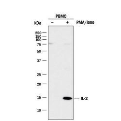
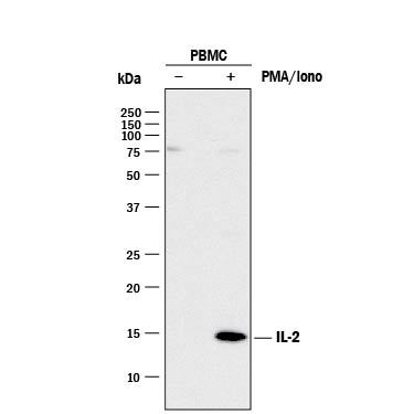
- Detection of Human IL-2 by Western Blot. Western blot shows lysates of monensin treated human peripheral blood mononuclear cells (PBMCs) with no additional treatment (-) or additionally treated (+) with 0.5 ug/mL calcium ionomycin (Iono) and 50 ng/mL PMA overnight. PVDF membrane was probed with 2 µg/mL of Goat Anti-Human/Rat IL-2 Antigen Affinity-purified Polyclonal Antibody (Catalog # AF-502-NA) followed by HRP-conjugated Anti-Goat IgG Secondary Antibody (Catalog # HAF017). A specific band was detected for IL-2 at approximately 14 kDa (as indicated). This experiment was conducted under reducing conditions and using Immunoblot Buffer Group 1.
Supportive validation
- Submitted by
- R&D Systems (provider)
- Main image
- Experimental details
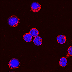
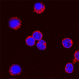
- IL-2 in Rat Splenocytes. IL-2 was detected in immersion fixed rat splenocytes stimulated with calcium ionomycin and PMA using Goat Anti-Human/Rat IL-2 Antigen Affinity-purified Polyclonal Antibody (Catalog # AF-502-NA) at 5 µg/mL for 3 hours at room temperature. Cells were stained using the NorthernLights™ 557-conjugated Anti-Goat IgG Secondary Antibody (red; Catalog # NL001) and counterstained with DAPI (blue). Specific staining was localized to cytoplasm. View our protocol for Fluorescent ICC Staining of Non-adherent Cells.
Supportive validation
- Submitted by
- R&D Systems (provider)
- Main image
- Experimental details
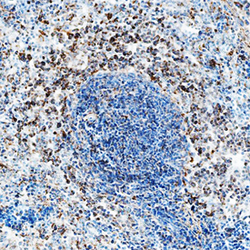
- IL-2 in Rat Spleen. IL-2 was detected in immersion fixed frozen sections of rat spleen using Goat Anti-Human/Rat IL-2 Antigen Affinity-purified Polyclonal Antibody (Catalog # AF-502-NA) at 5 µg/mL overnight at 4 °C. Tissue was stained using the Anti-Goat HRP-DAB Cell & Tissue Staining Kit (brown; Catalog # CTS008) and counterstained with hematoxylin (blue). Specific staining was localized to cytoplasm in lymphocytes. View our protocol for Chromogenic IHC Staining of Frozen Tissue Sections.

Supportive validation
- Submitted by
- R&D Systems (provider)
- Main image
- Experimental details
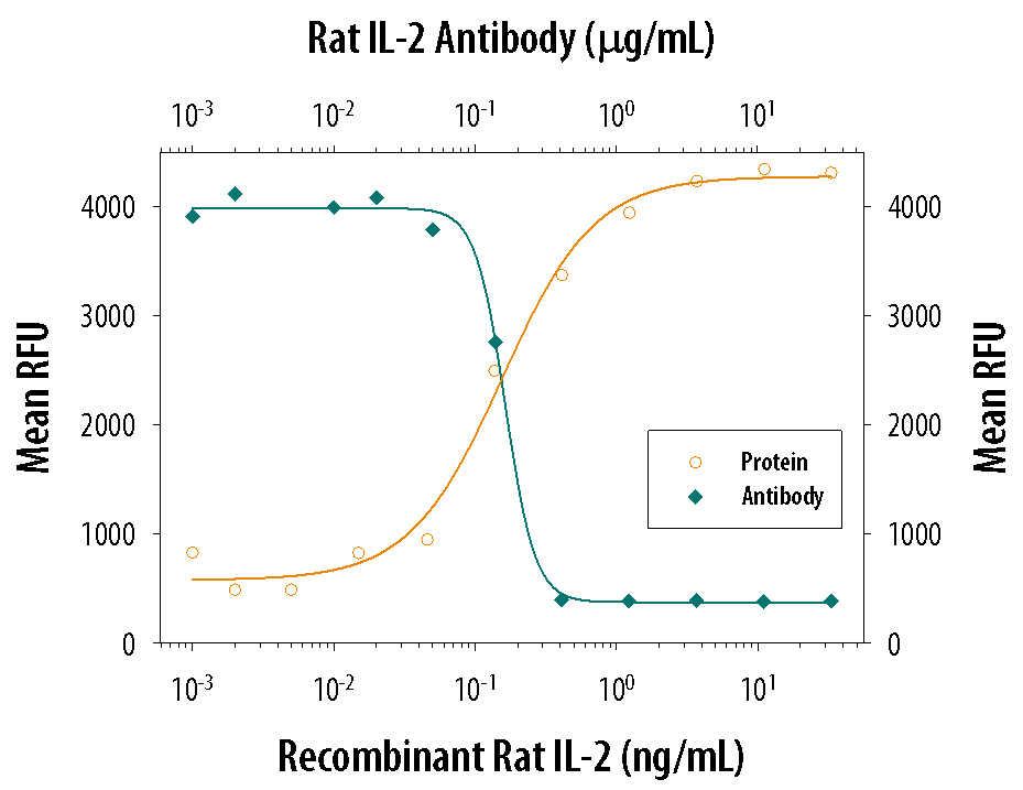
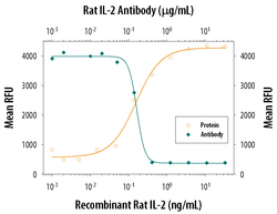
- Cell Proliferation Induced by IL-2 and Neutralization by Rat IL-2 Antibody. Recombinant Rat IL-2 (Catalog # 502-RL) stimulates proliferation in the CTLL-2 mouse cytotoxic T cell line in a dose-dependent manner (orange line). Proliferation elicited by Recombinant Rat IL-2 (2 ng/mL) is neutralized (green line) by increasing concentrations of Goat Anti-Human/Rat IL-2 Antigen Affinity-purified Polyclonal Antibody (Catalog # AF-502-NA). The ND50 is typically 0.15-0.75 µg/mL.