Explore
 Validate
 Learn
Learn Western blot
 ELISA
ELISA Immunocytochemistry
Antibody data
- Antibody Data
- Antigen structure
- References [2]
- Comments [0]
- Validations
- Immunocytochemistry [2]
- Other assay [2]
Submit
Validation data
Reference
Comment
Report error
- Product number
- MA5-15684 - Provider product page

- Provider
- Invitrogen Antibodies
- Product name
- Epo Monoclonal Antibody (4F11)
- Antibody type
- Monoclonal
- Antigen
- Purifed from natural sources
- Description
- MA5-15684 targets EPO in indirect ELISA, IF and WB applications and shows reactivity with Human samples. The MA5-15684 immunogen is purified recombinant fragment of human EPO expressed in E. Coli. MA5-15684 detects EPO which has a predicted molecular weight of approximately 21kDa.
- Reactivity
- Human
- Host
- Mouse
- Isotype
- IgG
- Antibody clone number
- 4F11
- Vial size
- 100 μL
- Concentration
- Conc. not determined
- Storage
- Store at 4°C short term. For long term storage, store at -20°C, avoiding freeze/thaw cycles.
Submitted references Chitosan and Hyaluronic Acid Nanoparticles as Vehicles of Epoetin Beta for Subconjunctival Ocular Delivery.
A therapeutic vascular conduit to support in vivo cell-secreted therapy.
Silva B, Gonçalves LM, Braz BS, Delgado E
Marine drugs 2022 Feb 18;20(2)
Marine drugs 2022 Feb 18;20(2)
A therapeutic vascular conduit to support in vivo cell-secreted therapy.
Han EX, Qian H, Jiang B, Figetakis M, Kosyakova N, Tellides G, Niklason LE, Chang WG
NPJ Regenerative medicine 2021 Jul 29;6(1):40
NPJ Regenerative medicine 2021 Jul 29;6(1):40
No comments: Submit comment
Supportive validation
- Submitted by
- Invitrogen Antibodies (provider)
- Main image

- Experimental details
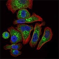
- Immunofluorescence analysis of GC7901 cells using EPO monoclonal antibody (Product # MA5-15684) (Green). Blue: DRAQ5 fluorescent DNA dye. Red: actin filaments have been labeled with phalloidin.

- Submitted by
- Invitrogen Antibodies (provider)
- Main image

- Experimental details
- Immunofluorescence analysis of GC7901 cells using EPO monoclonal antibody (Product # MA5-15684) (Green). Blue: DRAQ5 fluorescent DNA dye. Red: actin filaments have been labeled with phalloidin.
Supportive validation
- Submitted by
- Invitrogen Antibodies (provider)
- Main image
- Experimental details
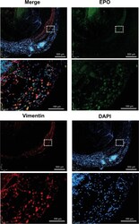
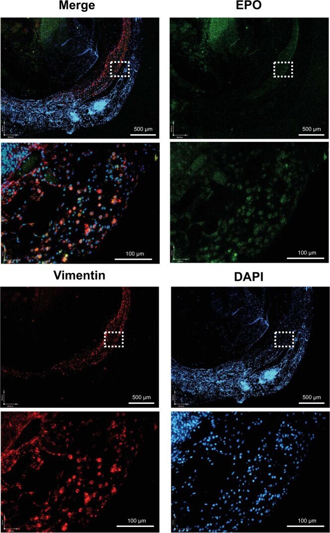
- Fig. 5 Immunofluorescent staining of EPO-TVC explant. TVC explant at 2 weeks stained with anti-EPO and anti-vimentin (FB marker) antibodies. Nuclei are stained with DAPI. TVC explant shows the outer layer of the TVC with EPO + FBs.
- Submitted by
- Invitrogen Antibodies (provider)
- Main image

- Experimental details
- Fig. 7 EPO, W6/32, and F4/80 staining of TVC explants. Implants from control and EPO-TVCs (2 weeks to 4 months) were stained for EPO by immunofluorescence and W6/32 (human HLA Class I) and F4/80 (macrophage marker) by immunohistochemistry. Control grafts were only examined at 3- and 4-month time points. Means with standard deviations are indicated for EPO 3- and 4-month time points where n = 2. For other time points, n = 1.