Explore
 Validate
 Learn
LearnPA5-76751
antibody from Invitrogen Antibodies
Targeting: GNAS
GNAS1, GNASXL, GPSA, NESP, NESP55, SCG6, SgVI
 Western blot
 Immunohistochemistry
Antibody data
- Antibody Data
- Antigen structure
- References [0]
- Comments [0]
- Validations
- Western blot [3]
Submit
Validation data
Reference
Comment
Report error
- Product number
- PA5-76751 - Provider product page

- Provider
- Invitrogen Antibodies
- Product name
- GNAS Polyclonal Antibody
- Antibody type
- Polyclonal
- Antigen
- Recombinant full-length protein
- Description
- The antibody was affinity-purified from rabbit antiserum by affinity-chromatography using epitope-specific immunogen and the purity is > 95% (by SDS-PAGE).
- Reactivity
- Human, Mouse, Rat
- Host
- Rabbit
- Isotype
- IgG
- Vial size
- 100 µL
- Concentration
- 1 mg/mL
- Storage
- Store at 4°C short term. For long term storage, store at -20°C, avoiding freeze/thaw cycles.
No comments: Submit comment
Supportive validation
- Submitted by
- Invitrogen Antibodies (provider)
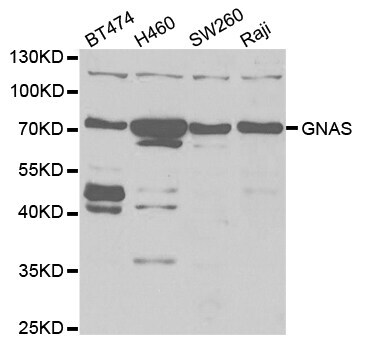
- Main image

- Experimental details
- Western blot analysis of GNAS. Samples were incubated with GNAS polyclonal antibody (Product # PA5-76751).
- Submitted by
- Invitrogen Antibodies (provider)
- Main image
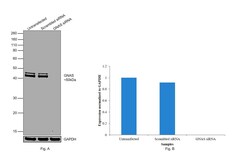
- Experimental details
- Knockdown of GNAS was achieved by transfecting Hep G2 with GNAS specific siRNAs (Silencer® select Products # s526399, s526398). Western blot analysis (Fig. a) was performed using whole cell extracts from the GNAS knockdown cells (Lane 3), non-specific scrambled siRNA transfected cells (Lane 2) and untransfected cells (Lane 1). The blot was probed with GNAS Polyclonal Antibody (Product # PA5-76751, 1:1000 dilution) and Goat anti-Rabbit IgG (H+L) Superclonal™ Recombinant Secondary Antibody, HRP (Product # A27036, 1:4000 dilution). Densitometric analysis of this western blot is shown in histogram (Fig. b). Loss of signal upon siRNA mediated knock down confirms that antibody is specific to GNAS.

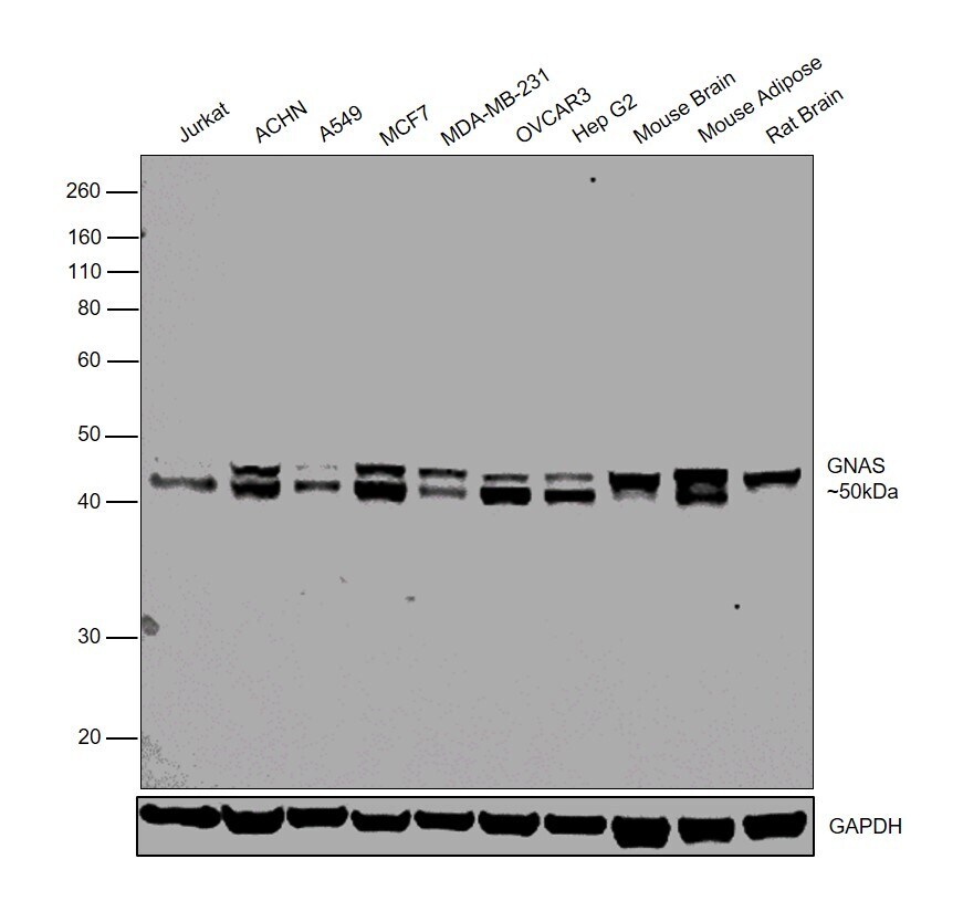
- Submitted by
- Invitrogen Antibodies (provider)
- Main image
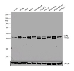
- Experimental details
- Western blot was performed using Anti-GNAS Polyclonal Antibody (Product # PA5-76751) and a 50 kDa band corresponding to GNAS was observed in all tested cell lines and tissue models. Whole cell lysates (30 µg lysate) of Jurkat (Lane 1), ACHN (Lane 2), A549 (Lane 3), MCF7 (Lane 4), MDA-MB-231 (Lane 5), OVCAR-3 (Lane 6), HepG2 (Lane 7), tissue lysates (30 µg lysate) of Mouse Brain (Lane 8), Mouse Adipose (Lane 9) and Rat Brain (Lane 10) were electrophoresed using NuPAGE® 10 % Bis-Tris gel (Product # NP0302BOX). Resolved proteins were then transferred onto a nitrocellulose membrane (Product # IB23001) by iBlot® 2 Dry Blotting System (Product # IB21001). The blot was probed with the primary antibody (1:1000 dilution) and detected by chemiluminescence with Goat anti-Rabbit IgG (H+L) Superclonal™ Recombinant Secondary Antibody, HRP (Product # A27036, 1:4000 dilution) using the iBright FL 1000 (Product # A32752). Chemiluminescent detection was performed using Novex® ECL Chemiluminescent Substrate Reagent Kit (Product # WP20005).