Explore
 Validate
 Learn
LearnPA5-19315
antibody from Invitrogen Antibodies
Targeting: GNAS
GNAS1, GNASXL, GPSA, NESP, NESP55, SCG6, SgVI
 Western blot
Antibody data
- Antibody Data
- Antigen structure
- References [2]
- Comments [0]
- Validations
- Western blot [3]
Submit
Validation data
Reference
Comment
Report error
- Product number
- PA5-19315 - Provider product page

- Provider
- Invitrogen Antibodies
- Product name
- GNAS Polyclonal Antibody
- Antibody type
- Polyclonal
- Antigen
- Synthetic peptide
- Description
- This antibody is predicted to react with bovine based on sequence homology.
- Reactivity
- Human, Mouse, Rat
- Host
- Goat
- Isotype
- IgG
- Vial size
- 100 µg
- Concentration
- 0.5 mg/mL
- Storage
- -20° C, Avoid Freeze/Thaw Cycles
Submitted references Human MC4R variants affect endocytosis, trafficking and dimerization revealing multiple cellular mechanisms involved in weight regulation.
G protein stoichiometry dictates biased agonism through distinct receptor-G protein partitioning.
Brouwers B, de Oliveira EM, Marti-Solano M, Monteiro FBF, Laurin SA, Keogh JM, Henning E, Bounds R, Daly CA, Houston S, Ayinampudi V, Wasiluk N, Clarke D, Plouffe B, Bouvier M, Babu MM, Farooqi IS, Mokrosiński J
Cell reports 2021 Mar 23;34(12):108862
Cell reports 2021 Mar 23;34(12):108862
G protein stoichiometry dictates biased agonism through distinct receptor-G protein partitioning.
Onfroy L, Galandrin S, Pontier SM, Seguelas MH, N'Guyen D, Sénard JM, Galés C
Scientific reports 2017 Aug 11;7(1):7885
Scientific reports 2017 Aug 11;7(1):7885
No comments: Submit comment
Supportive validation
- Submitted by
- Invitrogen Antibodies (provider)
- Main image
- Experimental details
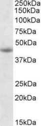
- Western Blot staining of Jurkat cell lysate using Product # PA5-19315 at a concentration of 1.0 µg/mL, the primary antibody incubation was 1 hour and the detection method was chemiluminescence.

- Submitted by
- Invitrogen Antibodies (provider)
- Main image
- Experimental details
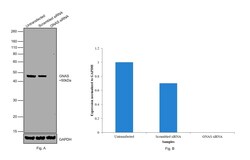
- Knockdown of GNAS was achieved by transfecting Hep G2 with GNAS specific siRNAs (Silencer® select Products # s526399, s526398). Western blot analysis (Fig. a) was performed using whole cell extracts from the GNAS knockdown cells (Lane 3), non-specific scrambled siRNA transfected cells (Lane 2) and untransfected cells (Lane 1). The blot was probed with GNAS Polyclonal Antibody (Product # PA5-19315, 0.3 µg/mL) and Rabbit anti-Goat IgG (H+L) Superclonal™ Recombinant Secondary Antibody, HRP (Product # A27014, 1:4000 dilution). Densitometric analysis of this western blot is shown in histogram (Fig. b). Loss of signal upon siRNA mediated knock down confirms that antibody is specific to GNAS.

- Submitted by
- Invitrogen Antibodies (provider)
- Main image
- Experimental details
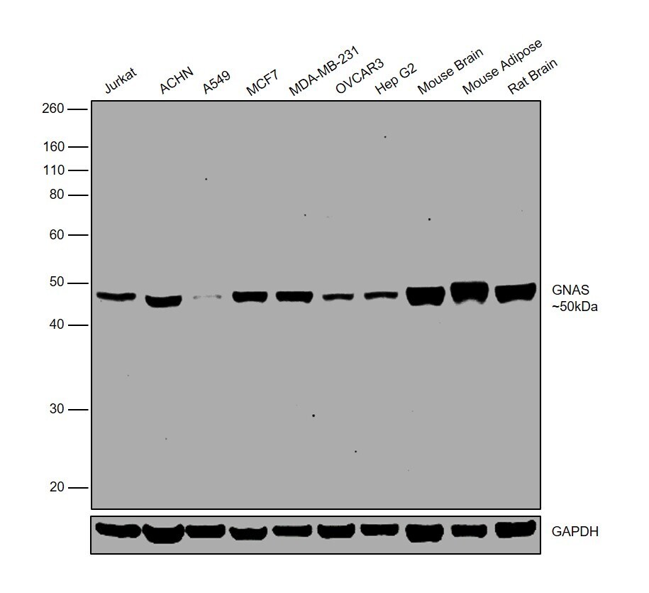
- Western blot was performed using Anti-GNAS Polyclonal Antibody (Product # PA5-19315) and a 50 kDa band corresponding to GNAS was observed in all tested cell lines and tissue models. Whole cell lysates (30 µg lysate) of Jurkat (Lane 1), ACHN (Lane 2), A549 (Lane 3), MCF7 (Lane 4), MDA-MB-231 (Lane 5), OVCAR-3 (Lane 6), HepG2 (Lane 7), tissue lysates (30 µg lysate) of Mouse Brain (Lane 8), Mouse Adipose (Lane 9) and Rat Brain (Lane 10) were electrophoresed using NuPAGE® 10 % Bis-Tris gel (Product # NP0302BOX). Resolved proteins were then transferred onto a nitrocellulose membrane (Product # IB23001) by iBlot® 2 Dry Blotting System (Product # IB21001). The blot was probed with the primary antibody (0.3 µg/mL) and detected by chemiluminescence with Rabbit anti-Goat IgG (H+L) Superclonal™ Recombinant Secondary Antibody, HRP (Product # A27014, 1:4000 dilution) using the iBright FL 1000 (Product # A32752). Chemiluminescent detection was performed using Novex® ECL Chemiluminescent Substrate Reagent Kit (Product # WP20005).