 Explore
 Validate
 Learn
LearnABIN498036
antibody from antibodies-online
Targeting: GNAS
GNAS1, GNASXL, GPSA, NESP, NESP55, SCG6, SgVI
 Western blot
 ELISA
ELISAAntibody data
- Antibody Data
- Antigen structure
- References [1]
- Comments [0]
- Validations
- Western blot [2]
Submit
Validation data
Reference
Comment
Report error
- Product number
- ABIN498036 - Provider product page

- Provider
- antibodies-online
- Product name
- anti-GNAS Complex Locus (GNAS) (Internal Region) antibody
- Antibody type
- Polyclonal
- Antigen
- Peptide with sequence from the internal region of the protein sequence according to NP_000507.1, NP_001070956.1, NP_001070957.1. Genename: GNAS
- Description
- Ammonium Sulphate Precipitation followed by Antigen Affinity Chromatography using the immunizing peptide
- Reactivity
- Human, Mouse, Rat, Bovine
- Host
- Goat
- Antigen sequence
C-QAARSNSDGEKATK- Epitope
- Internal Region
- Vial size
- 0.1 mg
- Concentration
- 0.5 mg/mL
- Storage
- Store undiluted at 2-8°C for one month or (in aliquots) at -20°C for longer.
- Handling
- Avoid repeated freezing and thawing.
Submitted references Prevalence of TSH receptor and Gsalpha mutations in 45 autonomously functioning thyroid nodules in Japan.
Nishihara E, Amino N, Maekawa K, Yoshida H, Ito M, Kubota S, Fukata S, Miyauchi A
Endocrine journal 2009;56(6):791-8
Endocrine journal 2009;56(6):791-8
No comments: Submit comment
Supportive validation
- Submitted by
- antibodies-online (provider)
- Main image
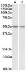
- Experimental details
- AP20123PU-N GNAS antibody staining of Mouse brain (Lane A) and Rat brain (Lane B) at 0.1 μg/ml (35μg protein in RIPA buffer). Primary incubated for 1 hour. Detected by western blot using chemiluminescence.

- Submitted by
- antibodies-online (provider)
- Main image
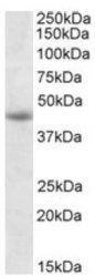
- Experimental details
- AP20123PU-N GNAS antibody staining of Jurkat lysate at 1 μg/ml (RIPA buffer, 35 μg total protein per lane). Primary incubated for 1 hour. Detected by western blot using chemiluminescence.
