 Explore
 Validate
 Learn
LearnMA5-35776
antibody from Invitrogen Antibodies
Targeting: SOCS2
CIS2, Cish2, SOCS-2, SSI-2, SSI2, STATI2
 Western blot
 ELISA
ELISAAntibody data
- Antibody Data
- Antigen structure
- References [0]
- Comments [0]
- Validations
- Western blot [2]
- Immunocytochemistry [3]
- Immunohistochemistry [2]
Submit
Validation data
Reference
Comment
Report error
- Product number
- MA5-35776 - Provider product page

- Provider
- Invitrogen Antibodies
- Product name
- SOCS2 Recombinant Rabbit Monoclonal Antibody (6D3F6)
- Antibody type
- Monoclonal
- Antigen
- Recombinant full-length protein
- Description
- Immunogen sequence: MTLRCLEPSG NGGEGTRSQW GTAGSAEEPS PQAARLAKAL RELGQTGWYW GSMTVNEAKE KLKEAPEGTF LIRDSSHSDY LLTISVKTSA GPTNLRIEYQ DGKFRLDSII CVKSKLKQFD SVVHLIDYYV QMCKDKRTGP EAPRNGTVHL YLTKPLYTSA PSLQHLCRLT INKCTGAIWG LPLPTRLKDY LEEYKFQV
- Reactivity
- Human, Mouse, Rat
- Host
- Rabbit
- Isotype
- IgG
- Antibody clone number
- 6D3F6
- Vial size
- 100 μL
- Concentration
- 2.0 mg/mL
- Storage
- -20°C, Avoid Freeze/Thaw Cycles
No comments: Submit comment
Supportive validation
- Submitted by
- Invitrogen Antibodies (provider)
- Main image

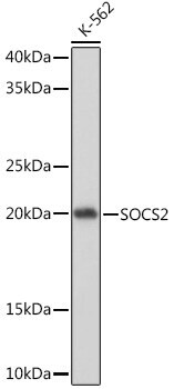
- Experimental details
- Western blot analysis of SOCS2 in extracts of K-562 cells. Samples were incubated with SOCS2 Monoclonal antibody (Product # MA5-35776) using a dilution of 1:1,000, followed by HRP Goat Anti-Rabbit IgG (H+L) at a dilution of 1:10,000. Lysates/proteins: 25 µg per lane. Blocking buffer: 3% nonfat dry milk in TBST. Detection: ECL Basic Kit. Exposure time: 1s.
- Submitted by
- Invitrogen Antibodies (provider)
- Main image
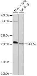
- Experimental details
- Western blot analysis of SOCS2 in extracts of various cell lines. Samples were incubated with SOCS2 Monoclonal antibody (Product # MA5-35776) using a dilution of 1:1,000, followed by HRP Goat Anti-Rabbit IgG (H+L) at a dilution of 1:10,000. Lysates/proteins: 25 µg per lane. Blocking buffer: 3% nonfat dry milk in TBST. Detection: ECL Enhanced Kit. Exposure time: 3 min.

Supportive validation
- Submitted by
- Invitrogen Antibodies (provider)
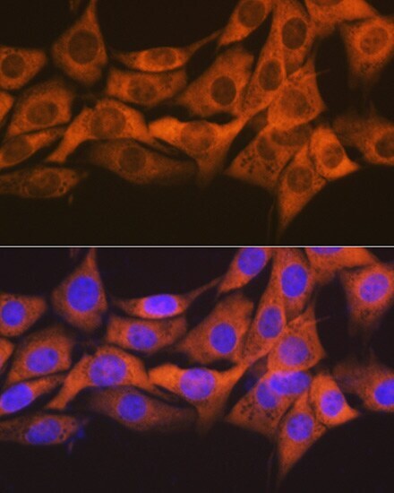
- Main image

- Experimental details
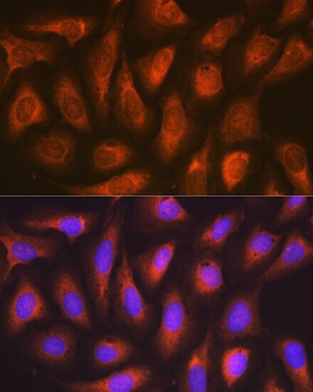
- Immunofluorescence analysis of NIH-3T3 cells using SOCS2 Monoclonal Antibody (Product # MA5-35776) diluted 1:100 (40x lens). Blue: DAPI for nuclear staining.

- Submitted by
- Invitrogen Antibodies (provider)
- Main image

- Experimental details
- Immunofluorescence analysis of SOCS2 in NIH/3T3 cells. Samples were incubated with SOCS2 Monoclonal antibody (Product # MA5-35776) using a dilution of 1:100 (40x lens). Blue: DAPI for nuclear staining.
- Submitted by
- Invitrogen Antibodies (provider)
- Main image
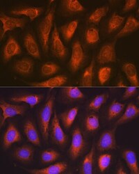
- Experimental details
- Immunofluorescence analysis of SOCS2 in U-2 OS cells. Samples were incubated with SOCS2 Monoclonal antibody (Product # MA5-35776) using a dilution of 1:100 (40x lens). Blue: DAPI for nuclear staining.
Supportive validation
- Submitted by
- Invitrogen Antibodies (provider)
- Main image

- Experimental details
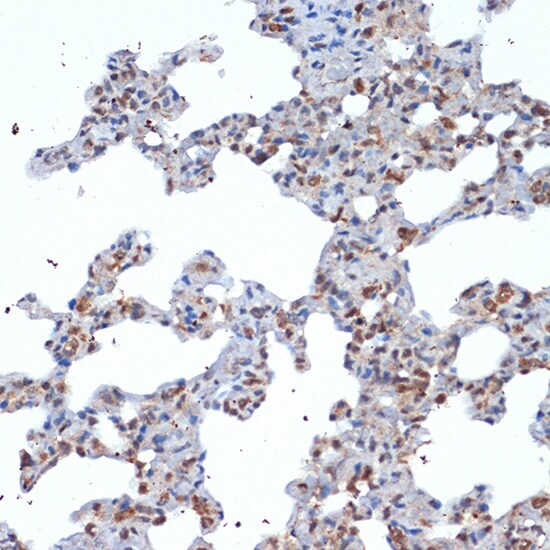
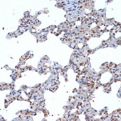
- Immunohistochemistry analysis of SOCS2 in paraffin-embedded rat lung. Samples were incubated with SOCS2 Monoclonal antibody (Product # MA5-35776) using a dilution of 1:100 (40x lens). Perform microwave antigen retrieval with 10 mM Tris/EDTA buffer pH 9.0 before commencing with IHC staining protocol.
- Submitted by
- Invitrogen Antibodies (provider)
- Main image
- Experimental details
- Immunohistochemistry analysis of SOCS2 in paraffin-embedded rat lung. Samples were incubated with SOCS2 Monoclonal antibody (Product # MA5-35776) using a dilution of 1:100 (40x lens). Perform microwave antigen retrieval with 10 mM Tris/EDTA buffer pH 9.0 before commencing with IHC staining protocol.
