 Explore
 Validate
 Learn
Learn Western blot
 Other assay
Antibody data
- Antibody Data
- Antigen structure
- References [0]
- Comments [0]
- Validations
- Other assay [4]
Submit
Validation data
Reference
Comment
Report error
- Product number
- MA1-19732 - Provider product page

- Provider
- Invitrogen Antibodies
- Product name
- PKA alpha Monoclonal Antibody (6D2.1)
- Antibody type
- Monoclonal
- Antigen
- Synthetic peptide
- Description
- This antibody reacts with human proteinkinase A (an intracellular antigen) catalytic (PKAc) alpha subunit, and weakly with PKAc gamma subunit (both around 40 kDa). The recognized epitope of PKAc alpha is identical between man, sheep, pig, ox and dog.
- Reactivity
- Human
- Host
- Mouse
- Isotype
- IgG
- Antibody clone number
- 6D2.1
- Vial size
- 100 μg
- Concentration
- 1 mg/mL
- Storage
- 4°C, do not freeze
No comments: Submit comment
Supportive validation
- Submitted by
- Invitrogen Antibodies (provider)
- Main image
- Experimental details
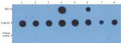
- Dot Blot analysis of GST and GST-fusion proteins using a nitrocellulose membrane (5 ng/spot); Lane 1: GST-Akt1, Lane 2: GST-Akt2, Lane 3: GST-Akt3, Lane 4: GST-PKAc alpha, Lane 5: GST-PKAc beta, Lane 6: GST-PKAc gamma, Lane 7: GST-MEK 1, Lane 8: GST.
- Submitted by
- Invitrogen Antibodies (provider)
- Main image
- Experimental details
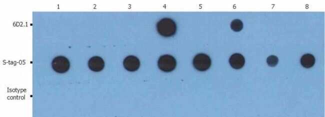
- Dot blot analysis of GST and GST-fusion proteins using anti-PKAc (6D2.1) and anti-GST (S-tag-05). The total amount of material spotted on the nitrocellulose membrane is 5 ng/spot. Lane 1: GST-Akt1; Lane 2: GST-Akt2; Lane 3: GST-Akt3; Lane 4: GST-PKAc alpha Monoclonal antibody (Product # MA1-19732); Lane 5: GST-PKAc beta; Lane 6: GST-PKAc gamma; Lane 7: GST-MEK 1; Lane 8: GST.

- Submitted by
- Invitrogen Antibodies (provider)
- Main image

- Experimental details
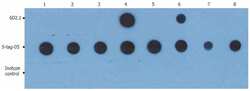
- Dot blot analysis of GST and GST-fusion proteins using anti-PKAc (6D2.1) and anti-GST (S-tag-05). The total amount of material spotted on the nitrocellulose membrane is 5 ng/spot. Lane 1: GST-Akt1; Lane 2: GST-Akt2; Lane 3: GST-Akt3; Lane 4: GST-PKAc alpha Monoclonal antibody (Product # MA1-19732); Lane 5: GST-PKAc beta; Lane 6: GST-PKAc gamma; Lane 7: GST-MEK 1; Lane 8: GST.

- Submitted by
- Invitrogen Antibodies (provider)
- Main image
- Experimental details
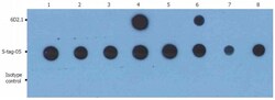
- Dot blot analysis of GST and GST-fusion proteins using anti-PKAc (6D2.1) and anti-GST (S-tag-05). The total amount of material spotted on the nitrocellulose membrane is 5 ng/spot. Lane 1: GST-Akt1; Lane 2: GST-Akt2; Lane 3: GST-Akt3; Lane 4: GST-PKAc alpha Monoclonal antibody (Product # MA1-19732); Lane 5: GST-PKAc beta; Lane 6: GST-PKAc gamma; Lane 7: GST-MEK 1; Lane 8: GST.
