Explore
 Validate
 Learn
Learn Western blot
 Immunocytochemistry
 Immunoprecipitation
Antibody data
- Antibody Data
- Antigen structure
- References [1]
- Comments [0]
- Validations
- Immunocytochemistry [2]
- Flow cytometry [2]
Submit
Validation data
Reference
Comment
Report error
- Product number
- PA5-17626 - Provider product page

- Provider
- Invitrogen Antibodies
- Product name
- PKA alpha Polyclonal Antibody
- Antibody type
- Polyclonal
- Antigen
- Synthetic peptide
- Description
- Antibodies to this protein (and modification) were previously sold as part of a Thermo Scientific Cellomics High Content Screening Kit. This replacement antibody is now recommended for researchers who need an antibody for high content cell based assays. It has been thoroughly tested and validated for cellular immunofluorescence (IF) applications. Further optimization including the selection of the most appropriate fluorescent Dylight conjugated secondary antibody may have to be performed for your high content assay. It is not recommended to aliquot this antibody.
- Reactivity
- Human, Mouse, Rat
- Host
- Rabbit
- Isotype
- IgG
- Vial size
- 100 μL
- Concentration
- 24 μg/mL
- Storage
- -20°C
Submitted references Preventive Effect of Limosilactobacillus fermentum SCHY34 on Lead Acetate-Induced Neurological Damage in SD Rats.
Long X, Wu H, Zhou Y, Wan Y, Kan X, Gong J, Zhao X
Frontiers in nutrition 2022;9:852012
Frontiers in nutrition 2022;9:852012
No comments: Submit comment
Supportive validation
- Submitted by
- Invitrogen Antibodies (provider)
- Main image
- Experimental details
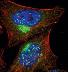
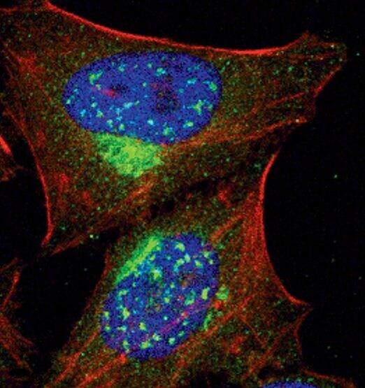
- Immunofluorescent analysis of PKA C-alpha in HeLa cells using a PKA C-alpha polyclonal antibody (Product # PA5-17626) (green). Actin filaments are labeled with a fluorescent red phalloidin. DNA is labeled using a fluorescent blue dye.

- Submitted by
- Invitrogen Antibodies (provider)
- Main image

- Experimental details
- Immunofluorescent analysis of PKA C-alpha in HeLa cells using a PKA C-alpha polyclonal antibody (Product # PA5-17626) (green). Actin filaments are labeled with a fluorescent red phalloidin. DNA is labeled using a fluorescent blue dye.
Supportive validation
- Submitted by
- Invitrogen Antibodies (provider)
- Main image
- Experimental details
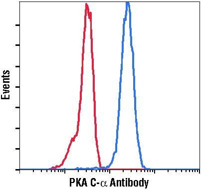
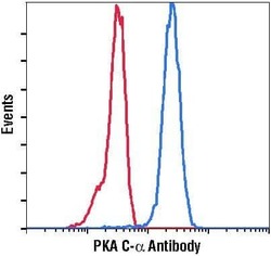
- Flow cytometric analysis of PKA C-alpha in HeLa cells using a PKA C-alpha polyclonal antibody (Product # PA5-17626) (blue) compared to a nonspecific negative control antibody (red).

- Submitted by
- Invitrogen Antibodies (provider)
- Main image

- Experimental details
- Flow cytometric analysis of PKA C-alpha in HeLa cells using a PKA C-alpha polyclonal antibody (Product # PA5-17626) (blue) compared to a nonspecific negative control antibody (red).