Explore
 Validate
 Learn
Learn Western blot
 Immunocytochemistry
Antibody data
- Antibody Data
- Antigen structure
- References [1]
- Comments [0]
- Validations
- Immunocytochemistry [5]
- Immunohistochemistry [4]
Submit
Validation data
Reference
Comment
Report error
- Product number
- PA5-22310 - Provider product page

- Provider
- Invitrogen Antibodies
- Product name
- HNF1A Polyclonal Antibody
- Antibody type
- Polyclonal
- Antigen
- Recombinant full-length protein
- Description
- Recommended positive controls: HepG2, Mouse liver, Rat liver. Predicted reactivity: Mouse (99%), Rat (99%), Xenopus laevis (83%), Pig (99%), Chicken (95%), Bovine (98%). Store product as a concentrated solution. Centrifuge briefly prior to opening the vial.
- Reactivity
- Human, Mouse, Rat
- Host
- Rabbit
- Isotype
- IgG
- Vial size
- 100 μL
- Concentration
- 1.27 mg/mL
- Storage
- Store at 4°C short term. For long term storage, store at -20°C, avoiding freeze/thaw cycles.
Submitted references Lack of TMEM27 expression is associated with postoperative progression of clinically localized conventional renal cell carcinoma.
Javorhazy A, Farkas N, Beothe T, Pusztai C, Szanto A, Kovacs G
Journal of cancer research and clinical oncology 2016 Sep;142(9):1947-53
Journal of cancer research and clinical oncology 2016 Sep;142(9):1947-53
No comments: Submit comment
Supportive validation
- Submitted by
- Invitrogen Antibodies (provider)
- Main image

- Experimental details
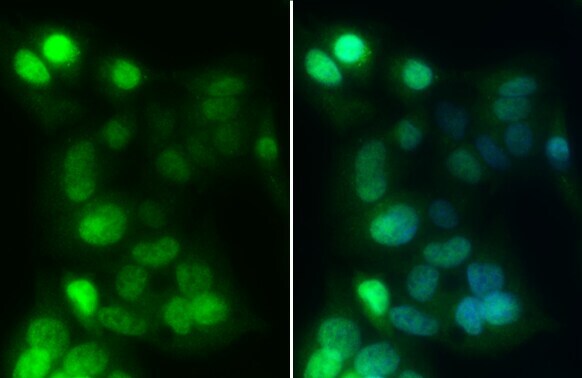
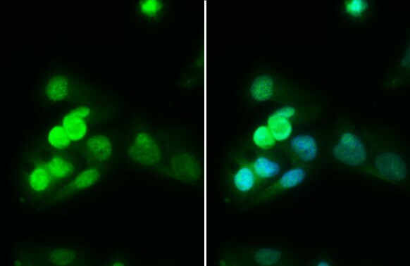
- HNF1A Polyclonal Antibody detects HNF1 alpha protein at nucleus by immunofluorescent analysis. Sample: HepG2 cells were fixed in 4% paraformaldehyde at RT for 15 min. Green: HNF1 alpha stained by HNF1A Polyclonal Antibody (Product # PA5-22310) diluted at 1:1,000. Blue: Fluoroshield with DAPI .

- Submitted by
- Invitrogen Antibodies (provider)
- Main image

- Experimental details
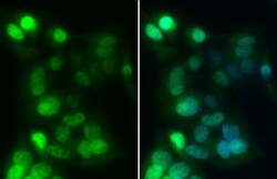
- Immunocytochemistry-Immunofluorescence analysis of HNF1A was performed in HepG2 cells fixed in 4% paraformaldehyde at RT for 15 min. Green: HNF1A Polyclonal Antibody (Product # PA5-22310) diluted at 1:1000. Blue: Hoechst 33342 staining.

- Submitted by
- Invitrogen Antibodies (provider)
- Main image

- Experimental details
- HNF1A Polyclonal Antibody detects HNF1 alpha protein at nucleus by immunofluorescent analysis. Sample: HepG2 cells were fixed in 4% paraformaldehyde at RT for 15 min. Green: HNF1 alpha stained by HNF1A Polyclonal Antibody (Product # PA5-22310) diluted at 1:1,000. Blue: Fluoroshield with DAPI .

- Submitted by
- Invitrogen Antibodies (provider)
- Main image
- Experimental details
- Immunocytochemistry-Immunofluorescence analysis of HNF1A was performed in HepG2 cells fixed in 4% paraformaldehyde at RT for 15 min. Green: HNF1A Polyclonal Antibody (Product # PA5-22310) diluted at 1:1000. Blue: Hoechst 33342 staining.
- Submitted by
- Invitrogen Antibodies (provider)
- Main image

- Experimental details
- HNF1A Polyclonal Antibody detects HNF1 alpha protein at nucleus by immunofluorescent analysis. Sample: HepG2 cells were fixed in 4% paraformaldehyde at RT for 15 min. Green: HNF1 alpha stained by HNF1A Polyclonal Antibody (Product # PA5-22310) diluted at 1:1,000. Blue: Fluoroshield with DAPI .
Supportive validation
- Submitted by
- Invitrogen Antibodies (provider)
- Main image
- Experimental details
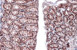
- Immunohistochemistry (Paraffin) analysis of HNF1A was performed in paraffin-embedded mouse colon tissue using HNF1A Polyclonal Antibody (Product # PA5-22310) at a dilution of 1:500. Antigen Retrieval: Citrate buffer, pH 6.0, 15 min.
- Submitted by
- Invitrogen Antibodies (provider)
- Main image
- Experimental details
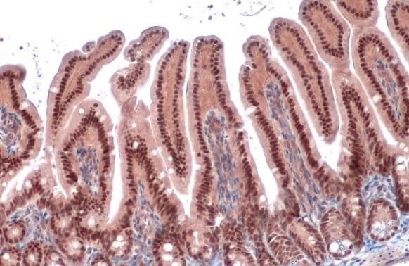
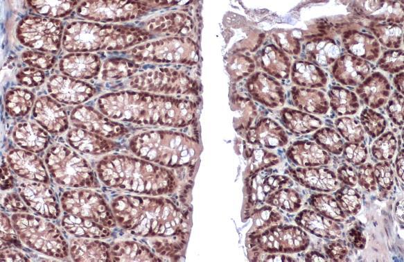
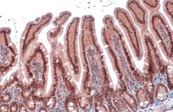
- HNF1A Polyclonal Antibody detects HNF1 alpha protein at nucleus by immunohistochemical analysis. Sample: Paraffin-embedded mouse intestine. HNF1 alpha stained by HNF1A Polyclonal Antibody (Product # PA5-22310) diluted at 1:500. Antigen Retrieval: Citrate buffer, pH 6.0, 15 min.
- Submitted by
- Invitrogen Antibodies (provider)
- Main image
- Experimental details
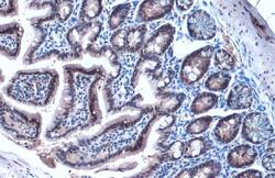
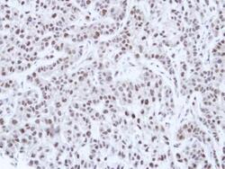
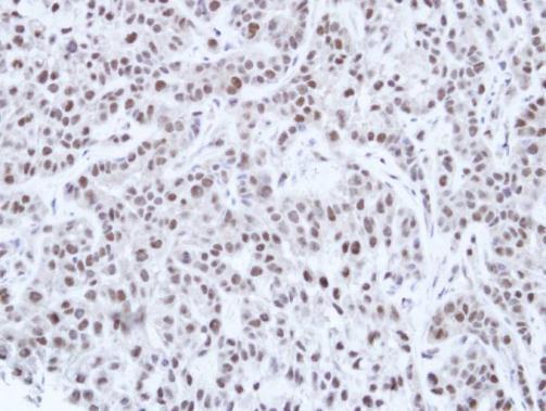
- Immunohistochemical analysis of paraffin-embedded A549 Xenograft, using HNF-1 alpha (Product # PA5-22310) antibody at 1:100 dilution. Antigen Retrieval: EDTA based buffer, pH 8.0, 15 min.
- Submitted by
- Invitrogen Antibodies (provider)
- Main image
- Experimental details
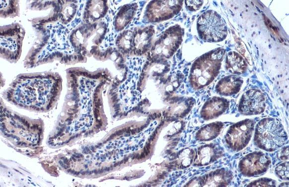
- Immunohistochemistry (Paraffin) analysis of HNF1A was performed in paraffin-embedded mouse intestine tissue using HNF1A Polyclonal Antibody (Product # PA5-22310) at a dilution of 1:500. Antigen Retrieval: Citrate buffer, pH 6.0, 15 min.