Explore
 Validate
 Learn
Learn Western blot
 Immunocytochemistry
Antibody data
- Antibody Data
- Antigen structure
- References [4]
- Comments [0]
- Validations
- Western blot [3]
- Immunoprecipitation [1]
- Immunohistochemistry [3]
Submit
Validation data
Reference
Comment
Report error
- Product number
- NBP1-33596 - Provider product page

- Provider
- Novus Biologicals
- Proper citation
- Novus Cat#NBP1-33596, RRID:AB_2248205
- Product name
- Rabbit Polyclonal HNF1 Antibody
- Antibody type
- Polyclonal
- Description
- Immunogen affinity purified. human HNF-1 alpha protein.
- Reactivity
- Human, Mouse, Rat
- Host
- Rabbit
- Isotype
- IgG
- Vial size
- 100 ul
- Storage
- Aliquot and store at -20C or -80C. Avoid freeze-thaw cycles.
Submitted references TGFβ Impairs HNF1α Functional Activity in Epithelial-to-Mesenchymal Transition Interfering With the Recruitment of CBP/p300 Acetyltransferases.
Effects of oolong tea on gene expression of gluconeogenic enzymes in the mouse liver and in rat hepatoma H4IIE cells.
Effects of (-)-epigallocatechin-3-O-gallate on expression of gluconeogenesis-related genes in the mouse duodenum.
Effects of (-)-epigallocatechin-3-O-gallate on expression of gluconeogenesis-related genes in the mouse duodenum.
Bisceglia F, Battistelli C, Noce V, Montaldo C, Zammataro A, Strippoli R, Tripodi M, Amicone L, Marchetti A
Frontiers in pharmacology 2019;10:942
Frontiers in pharmacology 2019;10:942
Effects of oolong tea on gene expression of gluconeogenic enzymes in the mouse liver and in rat hepatoma H4IIE cells.
Yasui K, Miyoshi N, Tababe H, Ishigami Y, Fukutomi R, Imai S, Isemura M
Journal of medicinal food 2011 Sep;14(9):930-8
Journal of medicinal food 2011 Sep;14(9):930-8
Effects of (-)-epigallocatechin-3-O-gallate on expression of gluconeogenesis-related genes in the mouse duodenum.
Yasui K, Tanabe H, Miyoshi N, Suzuki T, Goto S, Taguchi K, Ishigami Y, Paeng N, Fukutomi R, Imai S, Isemura M
Biomedical research (Tokyo, Japan) 2011 Oct;32(5):313-20
Biomedical research (Tokyo, Japan) 2011 Oct;32(5):313-20
No comments: Submit comment
Supportive validation
- Submitted by
- Novus Biologicals (provider)
- Main image
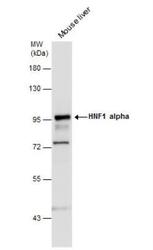
- Experimental details
- Western Blot: HNF1 Antibody [NBP1-33596] - Analysis. Mouse tissue extracts (50 Ug) was separated by 7.5% SDS-PAGE, and the membrane was blotted with HNF1 alpha antibody [N1N3] diluted at 1:2000.
- Submitted by
- Novus Biologicals (provider)
- Main image
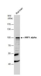
- Experimental details
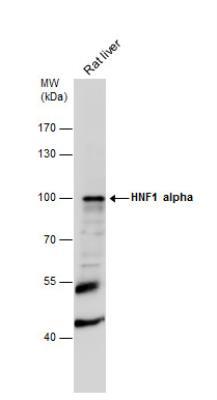
- Western Blot: HNF1 Antibody [NBP1-33596] - Rat tissue extracts (50 ug) was separated by 7.5% SDS-PAGE, and the membrane was blotted with HNF1 alpha antibody [N1N3] diluted at 1:1000.
- Submitted by
- Novus Biologicals (provider)
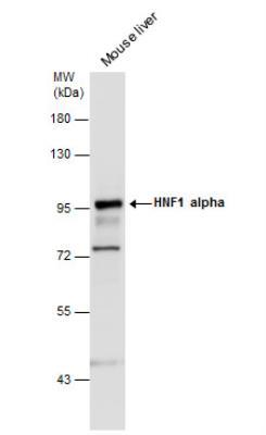
- Main image
- Experimental details
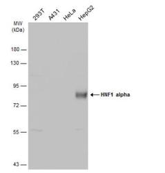
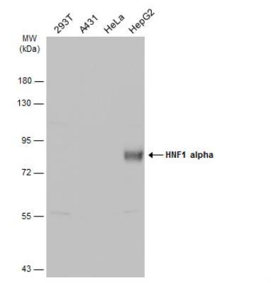
- Western Blot: HNF1 Antibody [NBP1-33596] - Various whole cell extracts (30 ug) were separated by 7.5% SDS-PAGE, and the membrane was blotted with HNF1 alpha antibody [N1N3] diluted at 1:5000. The HRP-conjugated anti-rabbit IgG antibody (NBP2-19301) was used to detect the primary antibody.
Supportive validation
- Submitted by
- Novus Biologicals (provider)
- Main image
- Experimental details
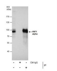
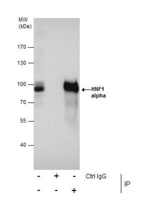
- Immunoprecipitation: HNF1 Antibody [NBP1-33596] - mmunoprecipitation of HNF1 alpha protein from HepG2 whole cell extracts using 5 ug of HNF1 alpha antibody [N1N3]. Western blot analysis was performed using HNF1 alpha antibody [N1N3]. EasyBlot anti-Rabbit IgG was used as a secondary reagent.
Supportive validation
- Submitted by
- Novus Biologicals (provider)
- Main image
- Experimental details
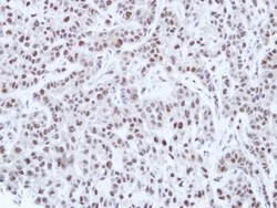
- Immunohistochemistry-Paraffin: HNF1 Antibody [NBP1-33596] - Paraffin-embedded A549 Xenograft, using antibody at 1:100 dilution.

- Submitted by
- Novus Biologicals (provider)
- Main image
- Experimental details
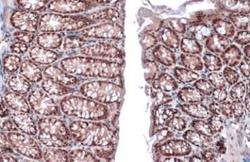
- Immunohistochemistry-Paraffin: HNF1 Antibody [NBP1-33596] - detects HNF1 alpha protein at nucleus by immunohistochemical analysis. Sample: Paraffin-embedded mouse colon. HNF1 alpha stained by HNF1 alpha antibody [N1N3] diluted at 1:500. Antigen Retrieval: Citrate buffer, pH 6.0, 15 min
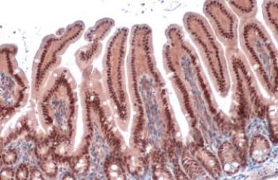
- Submitted by
- Novus Biologicals (provider)
- Main image
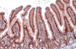
- Experimental details
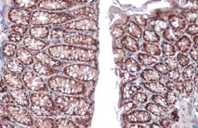
- Immunohistochemistry-Paraffin: HNF1 Antibody [NBP1-33596] - detects HNF1 alpha protein at nucleus by immunohistochemical analysis. Sample: Paraffin-embedded mouse intestine. HNF1 alpha stained by HNF1 alpha antibody [N1N3] diluted at 1:500. Antigen Retrieval: Citrate buffer, pH 6.0, 15 min