Explore
 Validate
 Learn
Learn Western blot
 Immunoprecipitation
Antibody data
- Antibody Data
- Antigen structure
- References [0]
- Comments [0]
- Validations
- Western blot [2]
Submit
Validation data
Reference
Comment
Report error
- Product number
- MA1-82085 - Provider product page

- Provider
- Invitrogen Antibodies
- Product name
- CDK4 Monoclonal Antibody (DCS-31.2)
- Antibody type
- Monoclonal
- Antigen
- Other
- Description
- Mouse anti CDK4 antibody clone DCS31.2 recognizes cyclin dependent kinase 4 (Cdk4), a ~34 kDa protein involved in the regulation of cell cycle progression between G1 and S.Mouse anti CDK4 antibody clone DCS31.2 does not cross react with other cdk's and does not co-precipitate the D type cyclins bound to cdk4.
- Reactivity
- Human, Mouse, Rat
- Host
- Mouse
- Isotype
- IgG
- Antibody clone number
- DCS-31.2
- Vial size
- 100 µg
- Concentration
- 1.0 mg/mL
- Storage
- Store at 4°C short term. For long term storage, store at -20°C, avoiding freeze/thaw cycles.
No comments: Submit comment
Supportive validation
- Submitted by
- Invitrogen Antibodies (provider)
- Main image

- Experimental details
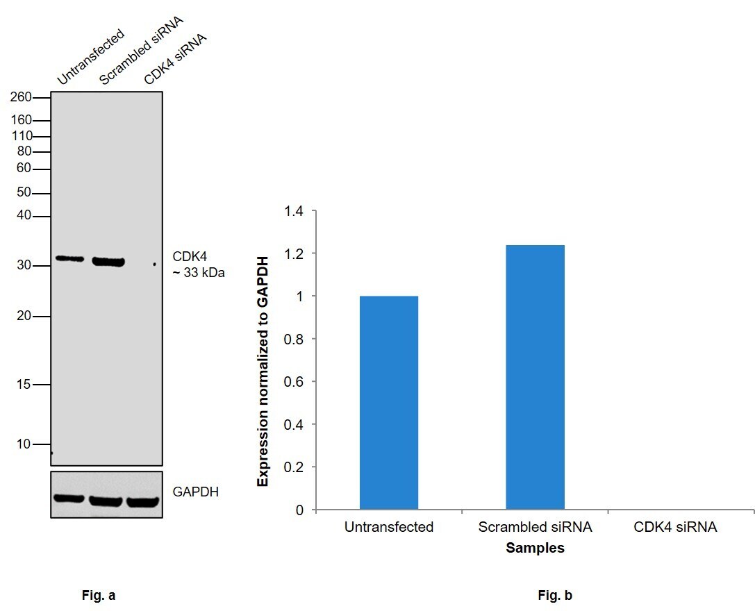
- Knockdown of CDK4 was achieved by transfecting HeLa with CDK4 specific siRNAs (Silencer® select Product # S2823, S2824). Western blot analysis (Fig. a) was performed using Whole cell extracts from the CDK4 knockdown cells (lane 3), non-targeting scrambled siRNA transfected cells (lane 2) and untransfected cells (lane 1). The blot was probed with CDK4 Monoclonal Antibody (DCS-31.2) (Product # MA1-82085, 1:1000 ) and Goat anti-Mouse IgG (H+L) Superclonal™ Recombinant Secondary Antibody, HRP (Product # A28177, 1:4000). Densitometric analysis of this western blot is shown in histogram (Fig. b). Decrease in signal upon siRNA mediated knock down confirms that antibody is specific to CDK4.
- Submitted by
- Invitrogen Antibodies (provider)
- Main image

- Experimental details
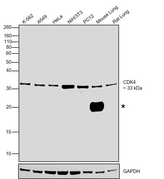
- Western blot was performed using Anti-CDK4 Monoclonal Antibody (DCS-31.2) (Product # MA1-82085) and a 33kDa band corresponding to CDK4 was observed across the cell lines tested and tissues tested along with uncharacterized band (*). Whole cell extracts (30 µg lysate) of K-562 (Lane 1), A549 (Lane 2), HeLa (Lane 3), NIH/3T3 (Lane 4), PC12 (Lane 5), Mouse Lung (Lane 6), Rat Lung (Lane 7) were electrophoresed using NuPAGE™ 4-12% Bis-Tris Protein Gel (Product # NP0322BOX). Resolved proteins were then transferred onto a Nitrocellulose membrane (Product # IB23002) by iBlot® 2 Dry Blotting System (Product # IB21001). The blot was probed with the primary antibody (1:1000) and detected by chemiluminescence with Goat anti-Mouse IgG (H+L) Superclonal™ Recombinant Secondary Antibody, HRP (Product # A28177, 1:2000) using the iBright FL 1000 (Product # A32752). Chemiluminescent detection was performed using Novex® ECL Chemiluminescent Substrate Reagent Kit (Product # WP20005).