Explore
 Validate
 Learn
Learn Western blot
Antibody data
- Antibody Data
- Antigen structure
- References [0]
- Comments [0]
- Validations
- Western blot [1]
- Immunocytochemistry [1]
- Immunohistochemistry [2]
Submit
Validation data
Reference
Comment
Report error
- Product number
- NBP2-32745 - Provider product page

- Provider
- Novus Biologicals
- Product name
- Rabbit Polyclonal CDK4 Antibody
- Antibody type
- Polyclonal
- Description
- Protein A purified.
- Reactivity
- Rat
- Host
- Rabbit
- Isotype
- IgG
- Vial size
- 400 ul
- Concentration
- 0.5 mg/ml
- Storage
- Store at 4C short term. Aliquot and store at -20C long term. Avoid freeze-thaw cycles.
No comments: Submit comment
Supportive validation
- Submitted by
- Novus Biologicals (provider)
- Main image

- Experimental details
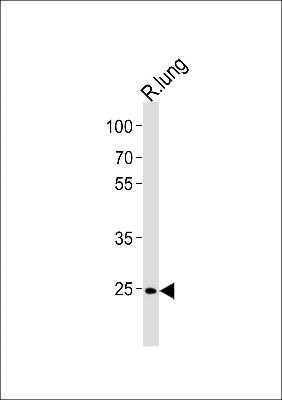
- Western Blot: CDk4 Antibody [NBP2-32745] - Rat Cdk4 Antibody (C-term) western blot analysis in rat lung tissue lysates (35ug/lane).This demonstrates the rat Cdk4 antibody detected the rat Cdk4 protein (arrow).
Supportive validation
- Submitted by
- Novus Biologicals (provider)
- Main image

- Experimental details
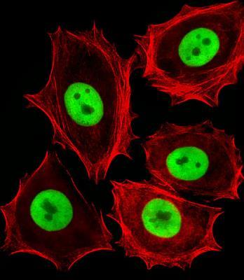
- Immunocytochemistry/Immunofluorescence: CDK4 Antibody [NBP2-32745] - MCF-7 cells stained with (Rat) Cdk4 Antibody.It was diluted at 1:25 dilution. An Alexa Fluor 488-conjugated goat anti-rabbit lgG at 1:400 dilution was used as the secondary antibody (green). Cytoplasmic actin was counterstained with Alexa Fluor 555 conjugated with Phalloidin (red).
Supportive validation
- Submitted by
- Novus Biologicals (provider)
- Main image
- Experimental details
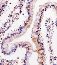
- Immunohistochemistry: CDK4 Antibody [NBP2-32745] - Analysis of paraffin-embedded rat small intestine section using Rat Cdk4 Antibody. It was diluted at 1:25 dilution. An undiluted biotinylated goat polyvalent antibody was used as the secondary, followed by DAB staining.

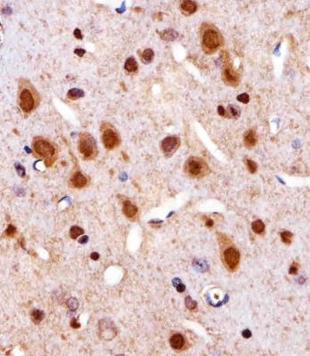
- Submitted by
- Novus Biologicals (provider)
- Main image

- Experimental details
- Immunohistochemistry: CDK4 Antibody [NBP2-32745] - Analysis of paraffin-embedded R. brain section using Cdk4 Antibody. It was diluted at 1:25 dilution. An undiluted biotinylated goat polyvalent antibody was used as the secondary, followed by DAB staining.