Explore
 Validate
 Learn
Learn Western blot
 Immunocytochemistry
Antibody data
- Antibody Data
- Antigen structure
- References [4]
- Comments [0]
- Validations
- Western blot [5]
- Immunohistochemistry [1]
Submit
Validation data
Reference
Comment
Report error
- Product number
- NBP1-31308 - Provider product page

- Provider
- Novus Biologicals
- Proper citation
- Novus Cat#NBP1-31308, RRID:AB_2078700
- Product name
- Rabbit Polyclonal CDK4 Antibody
- Antibody type
- Polyclonal
- Description
- Immunogen affinity purified.
- Reactivity
- Human, Mouse, Rat, Bovine, Feline
- Host
- Rabbit
- Isotype
- IgG
- Vial size
- 100 ul
- Storage
- Aliquot and store at -20C or -80C. Avoid freeze-thaw cycles.
Submitted references Prostaglandin F2α-PTGFR signaling promotes proliferation of endometrial epithelial cells of cattle through cell cycle regulation.
Proteins of the retinoblastoma pathway, FEN1 and MGMT are novel potential prognostic biomarkers in pancreatic adenocarcinoma.
Cell cycle S phase markers are expressed in cerebral neuron nuclei of cats infected by the Feline Panleukopenia Virus.
UBE2M-mediated p27(Kip1) degradation in gemcitabine cytotoxicity.
Fu C, Mao W, Gao R, Deng Y, Gao L, Wu J, Zhang S, Shen Y, Liu K, Li Q, Song X, Cao J, Liu B
Animal reproduction science 2020 Feb;213:106276
Animal reproduction science 2020 Feb;213:106276
Proteins of the retinoblastoma pathway, FEN1 and MGMT are novel potential prognostic biomarkers in pancreatic adenocarcinoma.
Isohookana J, Haapasaari KM, Soini Y, Leppänen J, Karihtala P
Pathology, research and practice 2018 Jun;214(6):840-847
Pathology, research and practice 2018 Jun;214(6):840-847
Cell cycle S phase markers are expressed in cerebral neuron nuclei of cats infected by the Feline Panleukopenia Virus.
Poncelet L, Garigliany M, Ando K, Franssen M, Desmecht D, Brion JP
Cell cycle (Georgetown, Tex.) 2016 Dec 16;15(24):3482-3489
Cell cycle (Georgetown, Tex.) 2016 Dec 16;15(24):3482-3489
UBE2M-mediated p27(Kip1) degradation in gemcitabine cytotoxicity.
Huang AM, Kao YT, Toh S, Lin PY, Chou CH, Hu HT, Lu CY, Liou JY, Chao SY, Hour TC, Pu YS
Biochemical pharmacology 2011 Jul 1;82(1):35-42
Biochemical pharmacology 2011 Jul 1;82(1):35-42
No comments: Submit comment
Supportive validation
- Submitted by
- Novus Biologicals (provider)
- Main image
- Experimental details
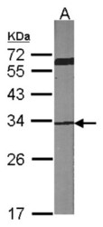
- Western Blot: Cdk4 Antibody [NBP1-31308] - Sample (30 ug of whole cell lysate) A:NIH-3T3 12% SDS PAGE; antibody diluted at 1:1000.

- Submitted by
- Novus Biologicals (provider)
- Main image
- Experimental details
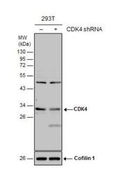
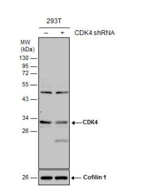
- Western Blot: CDK4 Antibody [NBP1-31308] - Non-transfected (-) and transfected (+) 293T whole cell extracts (30 ug) were separated by 12% SDS-PAGE, and the membrane was blotted with CDK4 antibody. HRP-conjugated anti-rabbit IgG antibody was used to detect the primary antibody.
- Submitted by
- Novus Biologicals (provider)
- Main image
- Experimental details
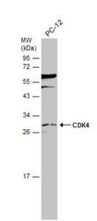
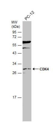
- Western Blot: CDK4 Antibody [NBP1-31308] - Whole cell extract (30 ug) was separated by 12% SDS-PAGE, and the membrane was blotted with CDK4 antibody diluted at 1:500. The HRP-conjugated anti-rabbit IgG antibody (NBP2-19301) was used to detect the primary antibody.
- Submitted by
- Novus Biologicals (provider)
- Main image
- Experimental details
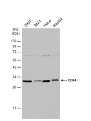
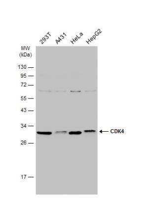
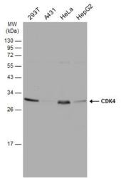
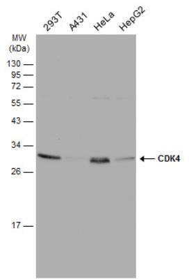
- Western Blot: CDK4 Antibody [NBP1-31308] - Various whole cell extracts (30 ug) were separated by 12% SDS-PAGE, and the membrane was blotted with CDK4 antibody diluted at 1:1000. The HRP-conjugated anti-rabbit IgG antibody (NBP2-19301) was used to detect the primary antibody.
- Submitted by
- Novus Biologicals (provider)
- Main image
- Experimental details
- Western Blot: CDK4 Antibody [NBP1-31308] - Various whole cell extracts (30 ug) were separated by 12% SDS-PAGE, and the membrane was blotted with CDK4 antibody diluted at 1:1000. The HRP-conjugated anti-rabbit IgG antibody (NBP2-19301) was used to detect the primary antibody.
Supportive validation
- Submitted by
- Novus Biologicals (provider)
- Main image

- Experimental details
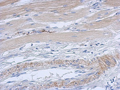
- Immunohistochemistry-Paraffin: Cdk4 Antibody [NBP1-31308] - Paraffin-embedded Gastric ca, using antibody at 1:500 dilution.