 Explore
 Validate
 Learn
Learn Western blot
Antibody data
- Antibody Data
- Antigen structure
- References [23]
- Comments [0]
- Validations
- Western blot [4]
- Immunocytochemistry [1]
- Flow cytometry [1]
- Other assay [1]
Submit
Validation data
Reference
Comment
Report error
- Product number
- MA5-13720 - Provider product page

- Provider
- Invitrogen Antibodies
- Product name
- CDK4 Antibody Cocktail
- Antibody type
- Monoclonal
- Antigen
- Purifed from natural sources
- Description
- MA5-13720 targets Cdk4 in IF, IP, and WB applications and shows reactivity with Human, mouse, and Rat samples.
- Antibody clone number
- DCS-31, DCS-35
- Concentration
- 0.2 mg/mL
Submitted references Mitochondrial ferritin, a new target for inhibiting neuronal tumor cell proliferation.
The APC/C activator Cdh1 regulates the G2/M transition during differentiation of placental trophoblast stem cells.
Phosphorylation of p27Kip1 regulates assembly and activation of cyclin D1-Cdk4.
Cyclic AMP inhibits the proliferation of thyroid carcinoma cell lines through regulation of CDK4 phosphorylation.
Regulation of keratinocyte proliferation in rats with deep, partial-thickness scald: modulation of cyclin D1-cyclin-dependent kinase 4 and histone H1 kinase activity of M-phase promoting factor.
Differential modification of p27Kip1 controls its cyclin D-cdk4 inhibitory activity.
Inhibition of prostate cancer growth by muscadine grape skin extract and resveratrol through distinct mechanisms.
Discovery of an oncogenic activity in p27Kip1 that causes stem cell expansion and a multiple tumor phenotype.
The LxCxE pRb interaction domain of cyclin D1 is dispensable for murine development.
Cyclin D1, cdk4, and Bim are involved in thrombin-induced apoptosis in cultured cortical neurons.
Requirement for CDK4 kinase function in breast cancer.
Cyclin D1-dependent kinase activity in murine development and mammary tumorigenesis.
p21Cip1 and p27Kip1 induce distinct cell cycle effects and differentiation programs in myeloid leukemia cells.
In vitro and in vivo pharmacokinetic-pharmacodynamic relationships for the trisubstituted aminopurine cyclin-dependent kinase inhibitors olomoucine, bohemine and CYC202.
p107 inhibits G1 to S phase progression by down-regulating expression of the F-box protein Skp2.
p53, p16 and cyclin D1: molecular determinants of radiotherapy treatment response in oral carcinoma.
The Cyclin-dependent kinase inhibitor CYC202 (R-roscovitine) inhibits retinoblastoma protein phosphorylation, causes loss of Cyclin D1, and activates the mitogen-activated protein kinase pathway.
Expression of angiotensin type II receptor downregulates Cdk4 synthesis and inhibits cell-cycle progression.
Regulation and role of p21 and p27 cyclin-dependent kinase inhibitors during hepatocyte differentiation and growth.
Interaction of Hsp90 with the nascent form of the mutant epidermal growth factor receptor EGFRvIII.
Reconstitution of cyclin D1-associated kinase activity drives terminally differentiated cells into the cell cycle.
Targeted disruption of CDK4 delays cell cycle entry with enhanced p27(Kip1) activity.
Targeted disruption of CDK4 delays cell cycle entry with enhanced p27(Kip1) activity.
Shi ZH, Shi FF, Wang YQ, Sheftel AD, Nie G, Zhao YS, You LH, Gou YJ, Duan XL, Zhao BL, Xu HM, Li CY, Chang YZ
Cellular and molecular life sciences : CMLS 2015 Mar;72(5):983-97
Cellular and molecular life sciences : CMLS 2015 Mar;72(5):983-97
The APC/C activator Cdh1 regulates the G2/M transition during differentiation of placental trophoblast stem cells.
Naoe H, Chiyoda T, Ishizawa J, Masuda K, Saya H, Kuninaka S
Biochemical and biophysical research communications 2013 Jan 11;430(2):757-62
Biochemical and biophysical research communications 2013 Jan 11;430(2):757-62
Phosphorylation of p27Kip1 regulates assembly and activation of cyclin D1-Cdk4.
Larrea MD, Liang J, Da Silva T, Hong F, Shao SH, Han K, Dumont D, Slingerland JM
Molecular and cellular biology 2008 Oct;28(20):6462-72
Molecular and cellular biology 2008 Oct;28(20):6462-72
Cyclic AMP inhibits the proliferation of thyroid carcinoma cell lines through regulation of CDK4 phosphorylation.
Rocha AS, Paternot S, Coulonval K, Dumont JE, Soares P, Roger PP
Molecular biology of the cell 2008 Nov;19(11):4814-25
Molecular biology of the cell 2008 Nov;19(11):4814-25
Regulation of keratinocyte proliferation in rats with deep, partial-thickness scald: modulation of cyclin D1-cyclin-dependent kinase 4 and histone H1 kinase activity of M-phase promoting factor.
Xie T, Niu Y, Ge K, Lu S
The Journal of surgical research 2008 Jun 1;147(1):9-14
The Journal of surgical research 2008 Jun 1;147(1):9-14
Differential modification of p27Kip1 controls its cyclin D-cdk4 inhibitory activity.
James MK, Ray A, Leznova D, Blain SW
Molecular and cellular biology 2008 Jan;28(1):498-510
Molecular and cellular biology 2008 Jan;28(1):498-510
Inhibition of prostate cancer growth by muscadine grape skin extract and resveratrol through distinct mechanisms.
Hudson TS, Hartle DK, Hursting SD, Nunez NP, Wang TT, Young HA, Arany P, Green JE
Cancer research 2007 Sep 1;67(17):8396-405
Cancer research 2007 Sep 1;67(17):8396-405
Discovery of an oncogenic activity in p27Kip1 that causes stem cell expansion and a multiple tumor phenotype.
Besson A, Hwang HC, Cicero S, Donovan SL, Gurian-West M, Johnson D, Clurman BE, Dyer MA, Roberts JM
Genes & development 2007 Jul 15;21(14):1731-46
Genes & development 2007 Jul 15;21(14):1731-46
The LxCxE pRb interaction domain of cyclin D1 is dispensable for murine development.
Landis MW, Brown NE, Baker GL, Shifrin A, Das M, Geng Y, Sicinski P, Hinds PW
Cancer research 2007 Aug 15;67(16):7613-20
Cancer research 2007 Aug 15;67(16):7613-20
Cyclin D1, cdk4, and Bim are involved in thrombin-induced apoptosis in cultured cortical neurons.
Rao HV, Thirumangalakudi L, Desmond P, Grammas P
Journal of neurochemistry 2007 Apr;101(2):498-505
Journal of neurochemistry 2007 Apr;101(2):498-505
Requirement for CDK4 kinase function in breast cancer.
Yu Q, Sicinska E, Geng Y, Ahnström M, Zagozdzon A, Kong Y, Gardner H, Kiyokawa H, Harris LN, Stål O, Sicinski P
Cancer cell 2006 Jan;9(1):23-32
Cancer cell 2006 Jan;9(1):23-32
Cyclin D1-dependent kinase activity in murine development and mammary tumorigenesis.
Landis MW, Pawlyk BS, Li T, Sicinski P, Hinds PW
Cancer cell 2006 Jan;9(1):13-22
Cancer cell 2006 Jan;9(1):13-22
p21Cip1 and p27Kip1 induce distinct cell cycle effects and differentiation programs in myeloid leukemia cells.
Muñoz-Alonso MJ, Acosta JC, Richard C, Delgado MD, Sedivy J, León J
The Journal of biological chemistry 2005 May 6;280(18):18120-9
The Journal of biological chemistry 2005 May 6;280(18):18120-9
In vitro and in vivo pharmacokinetic-pharmacodynamic relationships for the trisubstituted aminopurine cyclin-dependent kinase inhibitors olomoucine, bohemine and CYC202.
Raynaud FI, Whittaker SR, Fischer PM, McClue S, Walton MI, Barrie SE, Garrett MD, Rogers P, Clarke SJ, Kelland LR, Valenti M, Brunton L, Eccles S, Lane DP, Workman P
Clinical cancer research : an official journal of the American Association for Cancer Research 2005 Jul 1;11(13):4875-87
Clinical cancer research : an official journal of the American Association for Cancer Research 2005 Jul 1;11(13):4875-87
p107 inhibits G1 to S phase progression by down-regulating expression of the F-box protein Skp2.
Rodier G, Makris C, Coulombe P, Scime A, Nakayama K, Nakayama KI, Meloche S
The Journal of cell biology 2005 Jan 3;168(1):55-66
The Journal of cell biology 2005 Jan 3;168(1):55-66
p53, p16 and cyclin D1: molecular determinants of radiotherapy treatment response in oral carcinoma.
Jayasurya R, Francis G, Kannan S, Lekshminarayanan K, Nalinakumari KR, Abraham T, Abraham EK, Nair MK
International journal of cancer 2004 May 1;109(5):710-6
International journal of cancer 2004 May 1;109(5):710-6
The Cyclin-dependent kinase inhibitor CYC202 (R-roscovitine) inhibits retinoblastoma protein phosphorylation, causes loss of Cyclin D1, and activates the mitogen-activated protein kinase pathway.
Whittaker SR, Walton MI, Garrett MD, Workman P
Cancer research 2004 Jan 1;64(1):262-72
Cancer research 2004 Jan 1;64(1):262-72
Expression of angiotensin type II receptor downregulates Cdk4 synthesis and inhibits cell-cycle progression.
Gingras B, Rodier G, Giasson E, Coulombe P, Chassagne C, Meloche S
Oncogene 2003 May 1;22(17):2633-42
Oncogene 2003 May 1;22(17):2633-42
Regulation and role of p21 and p27 cyclin-dependent kinase inhibitors during hepatocyte differentiation and growth.
Ilyin GP, Glaise D, Gilot D, Baffet G, Guguen-Guillouzo C
American journal of physiology. Gastrointestinal and liver physiology 2003 Jul;285(1):G115-27
American journal of physiology. Gastrointestinal and liver physiology 2003 Jul;285(1):G115-27
Interaction of Hsp90 with the nascent form of the mutant epidermal growth factor receptor EGFRvIII.
Lavictoire SJ, Parolin DA, Klimowicz AC, Kelly JF, Lorimer IA
The Journal of biological chemistry 2003 Feb 14;278(7):5292-9
The Journal of biological chemistry 2003 Feb 14;278(7):5292-9
Reconstitution of cyclin D1-associated kinase activity drives terminally differentiated cells into the cell cycle.
Latella L, Sacco A, Pajalunga D, Tiainen M, Macera D, D'Angelo M, Felici A, Sacchi A, Crescenzi M
Molecular and cellular biology 2001 Aug;21(16):5631-43
Molecular and cellular biology 2001 Aug;21(16):5631-43
Targeted disruption of CDK4 delays cell cycle entry with enhanced p27(Kip1) activity.
Tsutsui T, Hesabi B, Moons DS, Pandolfi PP, Hansel KS, Koff A, Kiyokawa H
Molecular and cellular biology 1999 Oct;19(10):7011-9
Molecular and cellular biology 1999 Oct;19(10):7011-9
Targeted disruption of CDK4 delays cell cycle entry with enhanced p27(Kip1) activity.
Tsutsui T, Hesabi B, Moons DS, Pandolfi PP, Hansel KS, Koff A, Kiyokawa H
Molecular and cellular biology 1999 Oct;19(10):7011-9
Molecular and cellular biology 1999 Oct;19(10):7011-9
No comments: Submit comment
Supportive validation
- Submitted by
- Invitrogen Antibodies (provider)
- Main image
- Experimental details
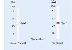
- Western blot of Cdk4 using Cdk4 Monoclonal Antibody (Product # MA5-13720) on MCF-7 Cells and PC12 Cells.

- Submitted by
- Invitrogen Antibodies (provider)
- Main image

- Experimental details
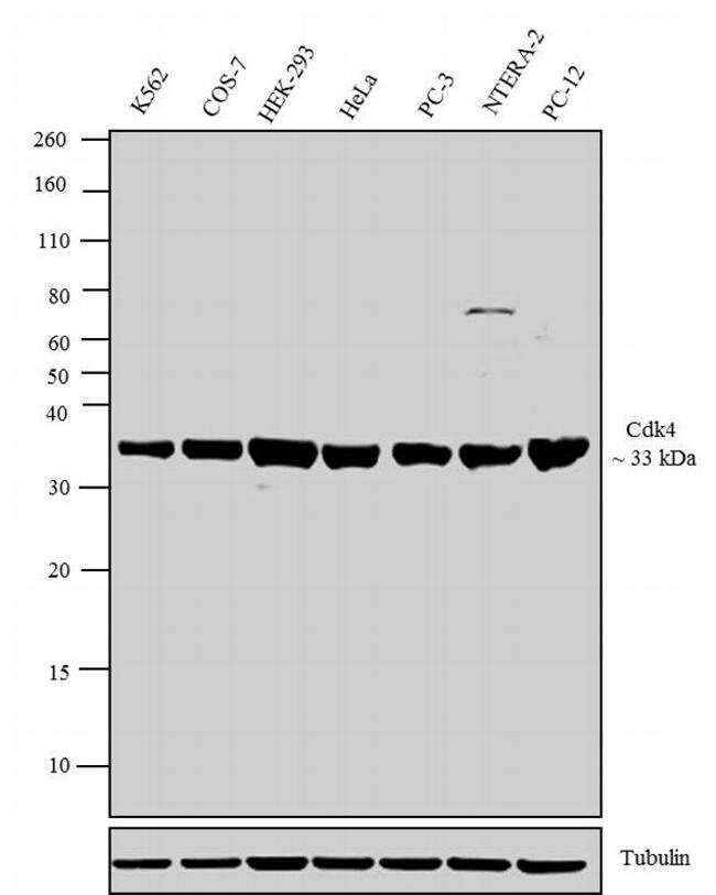
- Western blot analysis was performed on whole cell extracts (30 µg lysate) of K562 (Lane 1), COS-7 (Lane 2), HEK-293 (Lane 3), HeLa (Lane 4), PC-3 (Lane 5), NTERA-2 (Lane 6) and PC-12 (Lane 7). The blots were probed with Anti-Cdk4 Mouse Monoclonal Antibody (Product # MA5-13720, 1-3 µg/mL) and detected by chemiluminescence using Goat anti-Mouse IgG (H+L) Secondary Antibody, HRP conjugate (Product # 62-6520, 1:4000 dilution). A 33 kDa band corresponding to Cdk4 was observed across cell lines tested. Known quantity of protein samples were electrophoresed using Novex® NuPAGE® 12 % Bis-Tris gel (Product # NP0342BOX), XCell SureLock™ Electrophoresis System (Product # EI0002) and Novex® Sharp Pre-Stained Protein Standard (Product # LC5800). Resolved proteins were then transferred onto a nitrocellulose membrane with iBlot® 2 Dry Blotting System (Product # IB21001). The membrane was probed with the relevant primary and secondary Antibody following blocking with 5 % skimmed milk. Chemiluminescent detection was performed using Pierce™ ECL Western Blotting Substrate (Product # 32106).
- Submitted by
- Invitrogen Antibodies (provider)
- Main image
- Experimental details
- Knockout of CDK4 was achieved by CRISPR-Cas9 genome editing using LentiArray™ Lentiviral sgRNA (Product # A32042, Assay ID CRISPR1007917_LV) and LentiArray Cas9 Lentivirus (Product # A32064). Western blot analysis of CDK4 was performed by loading 30 µg of HeLa Wild Type (Lane 1), HeLa Cas9 (Lane 2) andHeLa CDK4 KO (Lane 3) whole cell extracts. The samples were electrophoresed using NuPAGE™ Novex™ 4-12% Bis-Tris Protein Gel (Product # NP0322BOX). Resolved proteins were then transferred onto a nitrocellulose membrane (Product # IB23001) by iBlot® 2 Dry Blotting System (Product # IB21001). The blot was probed with Anti-CDK4 Antibody Cocktail (Product # MA5-13720, 0.2 µg/mL dilution) and Goat anti-Mouse IgG (H+L) Superclonal™ Recombinant Secondary Antibody, HRP (Product # A28177, 1:5,000 dilution) using the iBright FL 1000 (Product # A32752). Chemiluminescent detection was performed using SuperSignal™ West Dura Extended Duration Substrate (Product # 34076). Loss of signal upon CRISPR mediated knockout (KO) using the LentiArray™ CRISPR product line confirms that antibody is specific to CDK4.

- Submitted by
- Invitrogen Antibodies (provider)
- Main image

- Experimental details
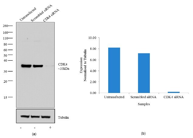
- Knockdown of CDK4 was achieved by transfecting HeLa cells with CDK4 specific validated siRNA (Silencer® select Product # s2823). Western blot analysis (Fig a) was performed using whole cell lysates from the CDK4 knock down cells (lane 3), non-specific scrambled siRNA transfected cells (lane 2) and untransfected cells (lane 1). The blots were probed with Anti-Cdk4 Mouse Monoclonal Antibody (Product # MA5-13720, 1-3 µg/mL) and Goat anti-Mouse IgG (H+L) Secondary Antibody, HRP conjugate (Product # 62-6520, 1:4000 dilution). Densitometric analysis of this western blot is shown in histogram(Fig b). Loss of signal upon siRNA mediated knock down confirms that antibody is specific to CDK4.
Supportive validation
- Submitted by
- Invitrogen Antibodies (provider)
- Main image
- Experimental details
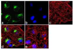
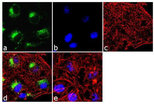
- Immunofluorescence analysis of Cdk4 was done on 70% confluent log phase HeLa cells. The cells were fixed with 4% paraformaldehyde for 10 minutes, permeabilized with 0.1% Triton™ X-100 for 10 minutes, and blocked with 1% BSA for 1 hour at room temperature. The cells were labeled with Cdk4 (DCS-31 + DCS-35) Mouse Monoclonal Antibody (Product # MA5-13720) at 2 µg/mL in 0.1% BSA and incubated for 3 hours at room temperature and then labeled with Goat anti-Mouse IgG (H+L) Superclonal™ Secondary Antibody, Alexa Fluor® 488 conjugate (Product # A28175) at a dilution of 1:2000 for 45 minutes at room temperature (Panel a: green). Nuclei (Panel b: blue) were stained with SlowFade® Gold Antifade Mountant with DAPI (Product # S36938). F-actin (Panel c: red) was stained with Alexa Fluor® 555 Rhodamine Phalloidin (Product # R415, 1:300). Panel d is a merged image peri-nuclear localization. Panel e is a no primary antibody control. The images were captured at 60X magnification.
Supportive validation
- Submitted by
- Invitrogen Antibodies (provider)
- Main image
- Experimental details
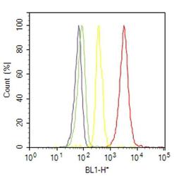
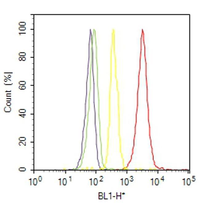
- Flow cytometry analysis of Cdk4 was done on K-562 cells. Cells were fixed with 70% ethanol for 10 minutes, permeabilized with 0.25% Triton™ X-100 for 20 minutes, and blocked with 5% BSA for 30 minutes at room temperature. Cells were labeled with Cdk4 Mouse Monoclonal Antibody (MA513720, red histogram) or with mouse isotype control (yellow histogram) at 3-5 ug/million cells in 2.5% BSA. After incubation at room temperature for 2 hours, the cells were labeled with Alexa Fluor® 488 Rabbit Anti-Mouse Secondary Antibody (A11059) at a dilution of 1:400 for 30 minutes at room temperature. The representative 10,000 cells were acquired and analyzed for each sample using an Attune® Acoustic Focusing Cytometer. The purple histogram represents unstained control cells and the green histogram represents no-primary-antibody control.
Supportive validation
- Submitted by
- Invitrogen Antibodies (provider)
- Main image
- Experimental details
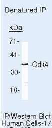
- Immunoprecipitation of Cdk4 using Cdk4 Monoclonal Antibody (Product # MA5-13720) on denatured Human LS174T Cells.
