Explore
 Validate
 Learn
Learn Western blot
 Immunocytochemistry
Antibody data
- Antibody Data
- Antigen structure
- References [1]
- Comments [0]
- Validations
- Immunocytochemistry [6]
- Immunohistochemistry [1]
- Other assay [2]
Submit
Validation data
Reference
Comment
Report error
- Product number
- PA5-27827 - Provider product page

- Provider
- Invitrogen Antibodies
- Product name
- CDK4 Polyclonal Antibody
- Antibody type
- Polyclonal
- Antigen
- Recombinant full-length protein
- Description
- Recommended positive controls: 293T, A431, HeLa, HepG2, NIH-3T3, PC-12. Predicted reactivity: Mouse (95%), Rat (94%), Pig (98%), Sheep (96%), Rhesus Monkey (100%), Bovine (96%).
- Reactivity
- Human, Mouse, Rat, Bovine
- Host
- Rabbit
- Isotype
- IgG
- Vial size
- 100 μL
- Concentration
- 0.82 mg/mL
- Storage
- Store at 4°C short term. For long term storage, store at -20°C, avoiding freeze/thaw cycles.
Submitted references Effects of lncRNA ANRIL-knockdown on the proliferation, apoptosis and cell cycle of gastric cancer cells.
Hu X, Lou T, Yuan C, Wang Y, Tu X, Wang Y, Zhang T
Oncology letters 2021 Aug;22(2):621
Oncology letters 2021 Aug;22(2):621
No comments: Submit comment
Supportive validation
- Submitted by
- Invitrogen Antibodies (provider)
- Main image
- Experimental details
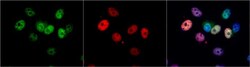
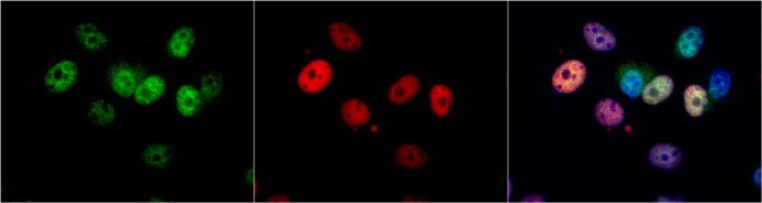
- Immunocytochemistry-Immunofluorescence analysis of CDK4 was performed in MCF7 cells fixed in 4% paraformaldehyde at RT for 15 min. Green: CDK4 Polyclonal Antibody (Product # PA5-27827) diluted at 1:1000. Red: p21 Cip1, a nucleus marker. Blue: Hoechst 33342 staining.
- Submitted by
- Invitrogen Antibodies (provider)
- Main image
- Experimental details
- Immunocytochemistry-Immunofluorescence analysis of CDK4 was performed in HeLa cells fixed in 4% paraformaldehyde at RT for 15 min. Green: CDK4 Polyclonal Antibody (Product # PA5-27827) diluted at 1:500. Red: phalloidin, a cytoskeleton marker. Scale bar = 10 µm.

- Submitted by
- Invitrogen Antibodies (provider)
- Main image
- Experimental details
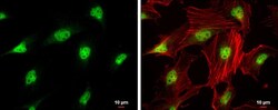
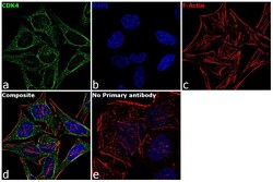
- Immunofluorescence analysis of CDK4 was performed using 70% confluent log phase HeLa cells. The cells were fixed with 4% paraformaldehyde for 10 minutes, permeabilized with 0.1% Triton™ X-100 for 15 minutes, and blocked with 1% BSA for 1 hour at room temperature. The cells were labeled with CDK4 Rabbit Polyclonal Antibody(Product # PA5-27827) at 5 µg/mL in 0.1% BSA, incubated at 4 degree Celsius overnight and then labeled with Goat anti-Rabbit IgG (H+L) Superclonal™ Secondary Antibody, Alexa Fluor® 488 conjugate (Product # A27034) at a dilution of 1:2000 for 45 minutes at room temperature (Panel a: green). Nuclei (Panel b: blue) were stained with SlowFade® Gold Antifade Mountant with DAPI (Product # S36938). F-actin (Panel c: red) was stained with Rhodamine Phalloidin (Product # R415, 1:300). Panel d represents the merged image showing cytoplasmic localization. Panel e represents control cells with no primary antibody to assess background. The images were captured at 60X magnification.
- Submitted by
- Invitrogen Antibodies (provider)
- Main image

- Experimental details
- Immunocytochemistry-Immunofluorescence analysis of CDK4 was performed in MCF7 cells fixed in 4% paraformaldehyde at RT for 15 min. Green: CDK4 Polyclonal Antibody (Product # PA5-27827) diluted at 1:1000. Red: p21 Cip1, a nucleus marker. Blue: Hoechst 33342 staining.

- Submitted by
- Invitrogen Antibodies (provider)
- Main image

- Experimental details
- Immunocytochemistry-Immunofluorescence analysis of CDK4 was performed in HeLa cells fixed in 4% paraformaldehyde at RT for 15 min. Green: CDK4 Polyclonal Antibody (Product # PA5-27827) diluted at 1:500. Red: phalloidin, a cytoskeleton marker. Scale bar = 10 µm.

- Submitted by
- Invitrogen Antibodies (provider)
- Main image
- Experimental details
- Immunofluorescence analysis of CDK4 was performed using 70% confluent log phase HeLa cells. The cells were fixed with 4% paraformaldehyde for 10 minutes, permeabilized with 0.1% Triton™ X-100 for 15 minutes, and blocked with 1% BSA for 1 hour at room temperature. The cells were labeled with CDK4 Rabbit Polyclonal Antibody(Product # PA5-27827) at 5 µg/mL in 0.1% BSA, incubated at 4 degree Celsius overnight and then labeled with Goat anti-Rabbit IgG (Heavy Chain) Superclonal™ Secondary Antibody, Alexa Fluor® 488 conjugate (Product # A27034) at a dilution of 1:2000 for 45 minutes at room temperature (Panel a: green). Nuclei (Panel b: blue) were stained with SlowFade® Gold Antifade Mountant with DAPI (Product # S36938). F-actin (Panel c: red) was stained with Rhodamine Phalloidin (Product # R415, 1:300). Panel d represents the merged image showing cytoplasmic localization. Panel e represents control cells with no primary antibody to assess background. The images were captured at 60X magnification.

Supportive validation
- Submitted by
- Invitrogen Antibodies (provider)
- Main image

- Experimental details
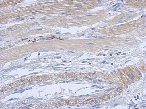
- Immunohistochemical analysis of paraffin-embedded human gastric cancer, using CDK4 (Product # PA5-27827) antibody at 1:500 dilution. Antigen Retrieval: EDTA based buffer, pH 8.0, 15 min.
Supportive validation
- Submitted by
- Invitrogen Antibodies (provider)
- Main image
- Experimental details
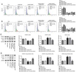
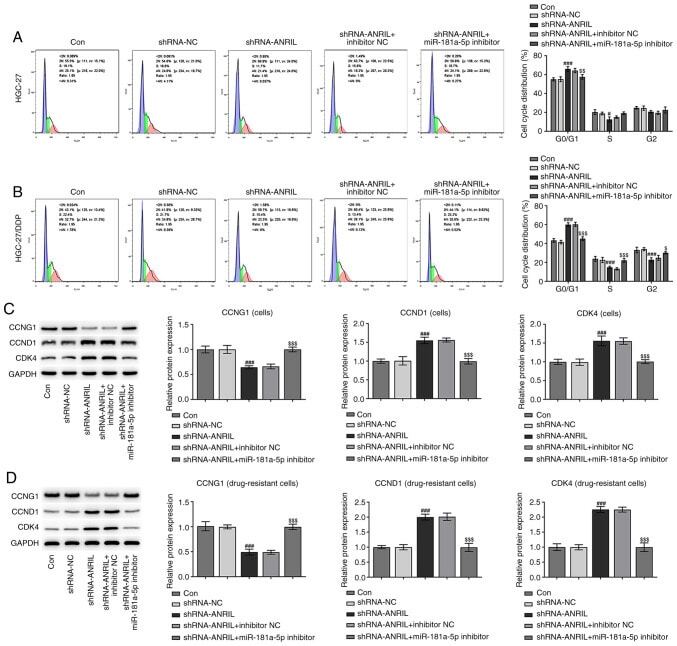
- Figure 6. Cell cycle distribution of HGC-27 and drug-resistant cell lines. Cell cycle distribution of (A) HGC-27 and (B) HGC-27 DDP-resistant cell lines was analyzed using flow cytometry. Expression of proteins related to the cell cycle in (C) HGC-27 and (D) HGC-27 DDP-resistant cell lines was analyzed using western blotting. # P
- Submitted by
- Invitrogen Antibodies (provider)
- Main image
- Experimental details
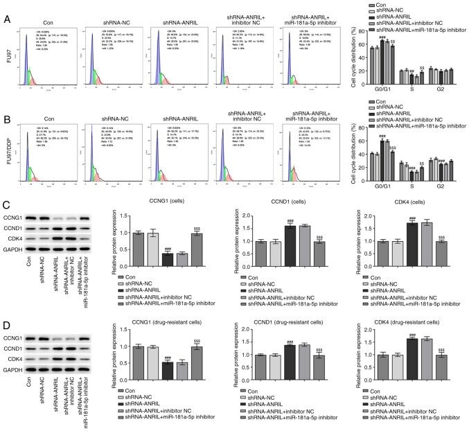
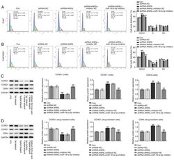
- Figure 7. Cell cycle distribution of FU97 and drug-resistant cell lines. Cell cycle distribution of (A) FU97 and (B) FU97 DDP-resistant cell lines was detected using flow cytometry. Expression of proteins related to the cell cycle in (C) FU97 and (D) FU97 DDP-resistant cell lines was analyzed using western blotting. ## P