Explore
 Validate
 Learn
Learn Flow cytometry
Antibody data
- Antibody Data
- Antigen structure
- References [5]
- Comments [0]
- Validations
- Flow cytometry [1]
- Other assay [1]
Submit
Validation data
Reference
Comment
Report error
- Product number
- 25-0059-42 - Provider product page

- Provider
- Invitrogen Antibodies
- Product name
- CD5 Monoclonal Antibody (UCHT2), PE-Cyanine7, eBioscience™
- Antibody type
- Monoclonal
- Antigen
- Other
- Description
- Description: The UCHT2 monoclonal antibody reacts with human CD5, a 67 kDa protein expressed by a majority of thymocytes and mature T cells and a subset of B cells. Signaling through the CD5 molecule activates T cells and binding of CD5 to its ligand on B cells, CD72, and plays an important role in T-B interaction and proliferation.\\\\The monoclonal antibody UCHT2 recognizes primate CD5. Applications Reported: This UCHT2 antibody has been reported for use in flow cytometric analysis. Applications Tested: This UCHT2 antibody has been pre-titrated and tested by flow cytometric analysis of normal human peripheral blood cells. This can be used at 5 µL (0.5 µg) per test. A test is defined as the amount (µg) of antibody that will stain a cell sample in a final volume of 100 µL. Cell number should be determined empirically but can range from 10^5 to 10^8 cells/test. Light sensitivity: This tandem dye is sensitive photo-induced oxidation. Please protect this vial and stained samples from light. Fixation: Samples can be stored in IC Fixation Buffer (Product # 00-822-49) (100 µL cell sample + 100 µL IC Fixation Buffer) or 1-step Fix/Lyse Solution (Product # 00-5333-54) for up to 3 days in the dark at 4°C with minimal impact on brightness and FRET efficiency/compensation. Some generalizations regarding fluorophore performance after fixation can be made, but clone specific performance should be determined empirically. Excitation: 488-561 nm; Emission: 775 nm; Laser: Blue Laser, Green Laser, Yellow-Green Laser. Filtration: 0.2 µm post-manufacturing filtered.
- Reactivity
- Human
- Host
- Mouse
- Isotype
- IgG
- Antibody clone number
- UCHT2
- Vial size
- 100 Tests
- Concentration
- 5 µL/Test
- Storage
- 4° C, store in dark, DO NOT FREEZE!
Submitted references In vitro OP9-DL1 co-culture and subsequent maturation in the presence of IL-21 generates tumor antigen-specific T cells with a favorable less-differentiated phenotype and enhanced functionality.
Targeted Disruption of TCF12 Reveals HEB as Essential in Human Mesodermal Specification and Hematopoiesis.
A Paradoxical Tumor-Suppressor Role for the Rac1 Exchange Factor Vav1 in T Cell Acute Lymphoblastic Leukemia.
Mimicking the tumour microenvironment of chronic lymphocytic leukaemia in vitro critically depends on the type of B-cell receptor stimulation.
A monoclonal antibody selection for immunohistochemical examination of lymphoid tissues from non-human primates.
Bonte S, de Munter S, Billiet L, Goetgeluk G, Ingels J, Jansen H, Pille M, de Cock L, Weening K, Taghon T, Leclercq G, Vandekerckhove B, Kerre T
Oncoimmunology 2021;10(1):1954800
Oncoimmunology 2021;10(1):1954800
Targeted Disruption of TCF12 Reveals HEB as Essential in Human Mesodermal Specification and Hematopoiesis.
Li Y, Brauer PM, Singh J, Xhiku S, Yoganathan K, Zúñiga-Pflücker JC, Anderson MK
Stem cell reports 2017 Sep 12;9(3):779-795
Stem cell reports 2017 Sep 12;9(3):779-795
A Paradoxical Tumor-Suppressor Role for the Rac1 Exchange Factor Vav1 in T Cell Acute Lymphoblastic Leukemia.
Robles-Valero J, Lorenzo-Martín LF, Menacho-Márquez M, Fernández-Pisonero I, Abad A, Camós M, Toribio ML, Espinosa L, Bigas A, Bustelo XR
Cancer cell 2017 Nov 13;32(5):608-623.e9
Cancer cell 2017 Nov 13;32(5):608-623.e9
Mimicking the tumour microenvironment of chronic lymphocytic leukaemia in vitro critically depends on the type of B-cell receptor stimulation.
Rombout A, Lust S, Offner F, Naessens E, Verhasselt B, Philippé J
British journal of cancer 2016 Mar 15;114(6):704-12
British journal of cancer 2016 Mar 15;114(6):704-12
A monoclonal antibody selection for immunohistochemical examination of lymphoid tissues from non-human primates.
Kap YS, van Meurs M, van Driel N, Koopman G, Melief MJ, Brok HP, Laman JD, 't Hart BA
The journal of histochemistry and cytochemistry : official journal of the Histochemistry Society 2009 Dec;57(12):1159-67
The journal of histochemistry and cytochemistry : official journal of the Histochemistry Society 2009 Dec;57(12):1159-67
No comments: Submit comment
Supportive validation
- Submitted by
- Invitrogen Antibodies (provider)
- Main image

- Experimental details
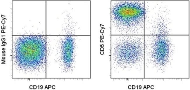
- Staining of normal human peripheral blood cells with Anti-Human CD19 APC (Product # 17-0199-42) and Mouse IgG1 kappa Isotype Control PE-Cyanine7 (Product # 25-4714-80) (left) or Anti-Human CD5 PE-Cyanine7 (right). Cells in the lymphocyte gate were used for analysis.
Supportive validation
- Submitted by
- Invitrogen Antibodies (provider)
- Main image
- Experimental details
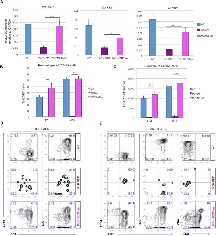
- Figure 7 HEBCan Rescues Hematopoiesis and T Cell Development in HEB -/- hESCs in OP9-DL4 Co-cultures (A) qRT-PCR analysis for the expression of hematopoietic genes in CD34 + cells sorted from WT, KO + GFP, and KO + HEBCan day-8 (d8) EBs. mRNA levels are shown relative to GAPDH. (B and C) Percentages (B) and numbers (C) of CD45 + cells in d12 and d18 OP9-DL4 co-cultures. (D and E) Flow-cytometric analysis of T cell development from WT, KO + GFP, and KO + HEBCan d8 EB-derived CD34 + cells at d12 (D) and d18 (E) of OP9-DL4 co-culture. Cells are gated on the CD45 + DAPI - population. Error bars represent mean +- SD (n = 3 independent experiments). * p < 0.05, ** p < 0.01, *** p < 0.005 by Student's t test. Plots in (B), (D), and (E) are representative of three independent experiments.