Explore
 Validate
 Learn
Learn Western blot
 Flow cytometry
Antibody data
- Antibody Data
- Antigen structure
- References [1]
- Comments [0]
- Validations
- Western blot [2]
- Immunocytochemistry [1]
- Other assay [1]
Submit
Validation data
Reference
Comment
Report error
- Product number
- AF1636 - Provider product page

- Provider
- R&D Systems
- Product name
- Human CD5 Antibody
- Antibody type
- Polyclonal
- Description
- Antigen Affinity-purified. Detects human CD5 in direct ELISAs and Western blots.
- Reactivity
- Human
- Host
- Goat
- Conjugate
- Unconjugated
- Antigen sequence
P06127- Isotype
- IgG
- Vial size
- 100 ug
- Concentration
- LYOPH
- Storage
- Use a manual defrost freezer and avoid repeated freeze-thaw cycles. 12 months from date of receipt, -20 to -70 °C as supplied. 1 month, 2 to 8 °C under sterile conditions after reconstitution. 6 months, -20 to -70 °C under sterile conditions after reconstitution.
Submitted references Unique cell surface expression of receptor tyrosine kinase ROR1 in human B-cell chronic lymphocytic leukemia.
Baskar S, Kwong KY, Hofer T, Levy JM, Kennedy MG, Lee E, Staudt LM, Wilson WH, Wiestner A, Rader C
Clinical cancer research : an official journal of the American Association for Cancer Research 2008 Jan 15;14(2):396-404
Clinical cancer research : an official journal of the American Association for Cancer Research 2008 Jan 15;14(2):396-404
No comments: Submit comment
Supportive validation
- Submitted by
- R&D Systems (provider)
- Main image
- Experimental details
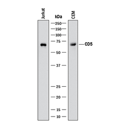
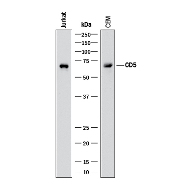
- Detection of Human CD5 by Western Blot. Western blot shows lysates of Jurkat human acute T cell leukemia cell line and CEM human T-lymphoblastoid cell line. PVDF membrane was probed with 0.5 µg/mL of Goat Anti-Human CD5 Antigen Affinity-purified Polyclonal Antibody (Catalog # AF1636) followed by HRP-conjugated Anti-Goat IgG Secondary Antibody (Catalog # HAF017). A specific band was detected for CD5 at approximately 67 kDa (as indicated). This experiment was conducted under reducing conditions and using Immunoblot Buffer Group 1.
- Submitted by
- R&D Systems (provider)
- Main image
- Experimental details
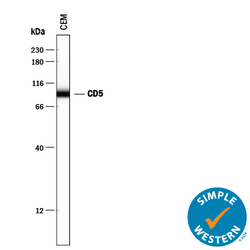
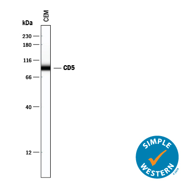
- Detection of Human CD5 by Simple WesternTM. Simple Western lane view shows lysates of CEM human T-lymphoblastoid cell line, loaded at 0.2 mg/mL. A specific band was detected for CD5 at approximately 93 kDa (as indicated) using 10 µg/mL of Goat Anti-Human CD5 Antigen Affinity-purified Polyclonal Antibody (Catalog # AF1636). This experiment was conducted under reducing conditions and using the 12-230 kDa separation system.
Supportive validation
- Submitted by
- R&D Systems (provider)
- Main image
- Experimental details
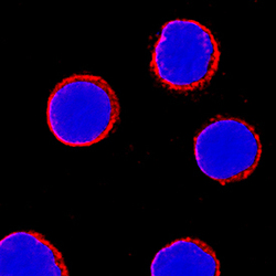
- CD5 in Human PBMCs. CD5 was detected in immersion fixed human peripheral blood mononuclear cells (PBMCs) using Goat Anti-Human CD5 Antigen Affinity-purified Polyclonal Antibody (Catalog # AF1636) at 5 µg/mL for 3 hours at room temperature. Cells were stained using the NorthernLights™ 557-conjugated Anti-Goat IgG Secondary Antibody (red; Catalog # NL001) and counterstained with DAPI (blue). Specific staining was localized to cell surfaces. View our protocol for Fluorescent ICC Staining of Non-adherent Cells.

Supportive validation
- Submitted by
- R&D Systems (provider)
- Main image
- Experimental details
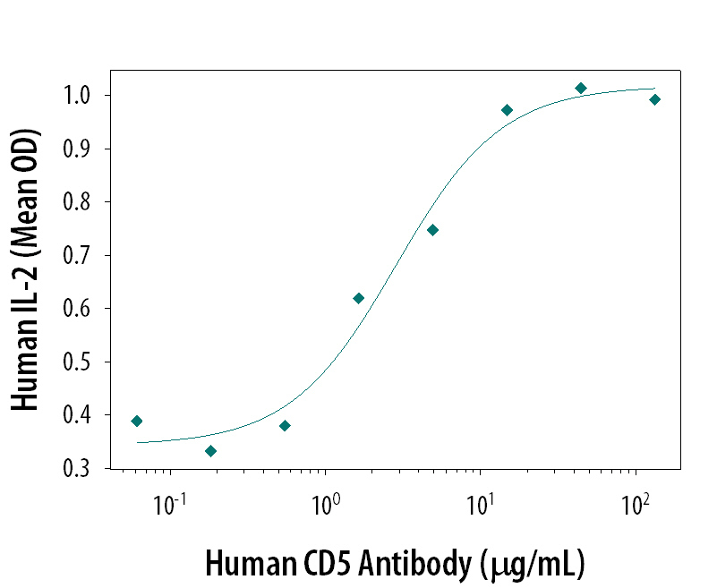
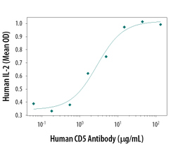
- Human CD5 Antibody Enhances IL-2 Secretion in Human T Cells. Human CD5 Antigen Affinity-purified Polyclonal Antibody enhances IL-2 secretion in human T cells in the presence of sub-optimal amounts of Human CD3 epsilon Monoclonal Antibody (Catalog # MAB100) and Human CD28 Monoclonal Antibody (Catalog # MAB342), in a dose-dependent manner, as measured using the Quantikine Human IL-2 ELISA Kit (Catalog # D2050). The ED50 for this effect is typically 2-8 μg/mL.