 Explore
 Validate
 Learn
Learn Western blot
Antibody data
- Antibody Data
- Antigen structure
- References [8]
- Comments [0]
- Validations
- Western blot [1]
Submit
Validation data
Reference
Comment
Report error
- Product number
- 100-401-249 - Provider product page

- Provider
- Rockland Immunochemicals, Inc.
- Proper citation
- Rockland Cat#100-401-249, RRID:AB_218037
- Product name
- Anti-CIITA aa 1-333 (RABBIT) Antibody - 100-401-249
- Antibody type
- Polyclonal
- Vial size
- 100 µl
Submitted references The Murine MHC Class II Super Enhancer IA/IE-SE Contains a Functionally Redundant CTCF-Binding Component and a Novel Element Critical for Maximal Expression.
EBNA2 driven enhancer switching at the CIITA-DEXI locus suppresses HLA class II gene expression during EBV infection of B-lymphocytes.
A super enhancer controls expression and chromatin architecture within the MHC class II locus.
Genome-wide CIITA-binding profile identifies sequence preferences that dictate function versus recruitment.
Regulation of acetylation at the major histocompatibility complex class II proximal promoter by the 19S proteasomal ATPase Sug1.
Influenza A virus abrogates IFN-gamma response in respiratory epithelial cells by disruption of the Jak/Stat pathway.
Hexim1 sequesters positive transcription elongation factor b from the class II transactivator on MHC class II promoters.
Self-association of class II transactivator correlates with its intracellular localization and transactivation.
Majumder P, Lee JT, Barwick BG, Patterson DG, Bally APR, Scharer CD, Boss JM
Journal of immunology (Baltimore, Md. : 1950) 2021 May 1;206(9):2221-2232
Journal of immunology (Baltimore, Md. : 1950) 2021 May 1;206(9):2221-2232
EBNA2 driven enhancer switching at the CIITA-DEXI locus suppresses HLA class II gene expression during EBV infection of B-lymphocytes.
Su C, Lu F, Soldan SS, Lamontagne RJ, Tang HY, Napoletani G, Farrell PJ, Tempera I, Kossenkov AV, Lieberman PM
PLoS pathogens 2021 Aug;17(8):e1009834
PLoS pathogens 2021 Aug;17(8):e1009834
A super enhancer controls expression and chromatin architecture within the MHC class II locus.
Majumder P, Lee JT, Rahmberg AR, Kumar G, Mi T, Scharer CD, Boss JM
The Journal of experimental medicine 2020 Feb 3;217(2)
The Journal of experimental medicine 2020 Feb 3;217(2)
Genome-wide CIITA-binding profile identifies sequence preferences that dictate function versus recruitment.
Scharer CD, Choi NM, Barwick BG, Majumder P, Lohsen S, Boss JM
Nucleic acids research 2015 Mar 31;43(6):3128-42
Nucleic acids research 2015 Mar 31;43(6):3128-42
Regulation of acetylation at the major histocompatibility complex class II proximal promoter by the 19S proteasomal ATPase Sug1.
Koues OI, Dudley RK, Truax AD, Gerhardt D, Bhat KP, McNeal S, Greer SF
Molecular and cellular biology 2008 Oct;28(19):5837-50
Molecular and cellular biology 2008 Oct;28(19):5837-50
Influenza A virus abrogates IFN-gamma response in respiratory epithelial cells by disruption of the Jak/Stat pathway.
Uetani K, Hiroi M, Meguro T, Ogawa H, Kamisako T, Ohmori Y, Erzurum SC
European journal of immunology 2008 Jun;38(6):1559-73
European journal of immunology 2008 Jun;38(6):1559-73
Hexim1 sequesters positive transcription elongation factor b from the class II transactivator on MHC class II promoters.
Kohoutek J, Blazek D, Peterlin BM
Proceedings of the National Academy of Sciences of the United States of America 2006 Nov 14;103(46):17349-54
Proceedings of the National Academy of Sciences of the United States of America 2006 Nov 14;103(46):17349-54
Self-association of class II transactivator correlates with its intracellular localization and transactivation.
Kretsovali A, Spilianakis C, Dimakopoulos A, Makatounakis T, Papamatheakis J
The Journal of biological chemistry 2001 Aug 24;276(34):32191-7
The Journal of biological chemistry 2001 Aug 24;276(34):32191-7
No comments: Submit comment
Supportive validation
- Submitted by
- Rockland Immunochemicals, Inc. (provider)
- Main image
- Experimental details
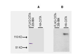
- Western blot of Anti-CIITA (1-333) antibody, generated by immunization with bacterially produced FLAG-CIITA aa 1-333, was tested by western blot against lysates of Cos-7 cells after transient transfection, separately, with pcDNA3-FLAG-CIITA-336-884 and pcDNA3-HA-CIITA. For transfection, Fugene 6 (Roche) was used according to the manufacturer's instructions for a 6-well plate format. Cells were lysed 24 h post-transfection in 200 uL of 1x SDS-sample buffer, heated at 96C for 5', and vortexed for 30 sec. Samples (10 uL each) were separated on a 12% SDS-PAGE gel and transferred to PVDF (Millipore) followed by blocking for 45' using TTBS supplemented with 5% non-fat dry milk. All incubations were performed at room temperature. In panel A, both samples on PVDF were incubated with 10 ug/mL mouse anti-FLAG antibody (Sigma) for 45'. After 5X washes with TTBS, reaction with ALP rabbit anti-mouse IgG at 200 ng/mL proceeded for 45' following again by washing as before. The blot was developed using BCIP/NBT. This blot demonstrates that FLAG-CIITA-336-842 was successfully over-expressed in the Cos-7 cells. In panel B, both samples on PVDF were incubated with a 1:500 dilution of Rockland's anti-CIITA (1-333) for 45'. After 5X washes with TTBS, reaction with HRP goat anti-rabbit IgG at 10 ng/mL proceeded for 45' following again by washing as before. The membrane was covered with Pico West Substrate solution (Pierce) for 5' and was then placed between the two layers of a standard sheet protector. Kodak O-MAT film was exposed to the blot for 30 sec and was immediately developed. The lane containing the lysate of pcDNA3-HA-CIITA transfected cells contains a single band at ~130 kD molecular weight, whereas the lane containing lysate from pcDNA3-FLAG-CIITA-336-842 transfected cells shows no reactivity. This blot demonstrates that anti-CIITA (1-333) is specific for amino acids 1-333 of CIITA and that the antibody is not cross reactive with the FLAG portion of the immunogen.
- Validation comment
- Western Blot
