Explore
 Validate
 Learn
Learn Flow cytometry
Antibody data
- Antibody Data
- Antigen structure
- References [5]
- Comments [0]
- Validations
- Flow cytometry [1]
- Other assay [2]
Submit
Validation data
Reference
Comment
Report error
- Product number
- 12-0016-42 - Provider product page

- Provider
- Invitrogen Antibodies
- Product name
- CD1d Monoclonal Antibody (51.1), PE, eBioscience™
- Antibody type
- Monoclonal
- Antigen
- Other
- Description
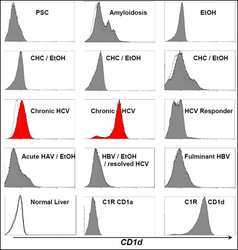
- Description: The monoclonal antibody 51.1 reacts with human CD1d, a member of the CD1 family with similarity to the non-polymorphic MHC Class I-like molecules. CD1d is a highly conserved single transmembrane receptor of the Immunoglobulin Superfamily. CD1d can associate with beta-microglobulin another feature showing similarity to MHC class I molecules, but can also exist as a nonglycosylated protein not in association with beta microglobulin. This suggests different control mechanisms for presenting glycolipid containing molecules to CD1d reactive NKT cells. Expression of CD1d is found on B cells of the periphery, in resting monocytes and cortical thymocytes. On intestinal epithelial cells (IEC) expression is polarized. Expression can also be found at low levels intracellularly in hepatocytes. In HCV (hepatitis C virus) livers, CD1d is highly expressed compared to normal controls.
- Conjugate
- Yellow dye
- Antibody clone number
- 51.1
- Concentration
- 5 µL/Test
Submitted references Patients with Tuberculosis Have a Dysfunctional Circulating B-Cell Compartment, Which Normalizes following Successful Treatment.
Expression of CD1a and Type-1 Polarization Are Dissociated in Human Monocyte-Derived Dendritic Cells.
CD1d levels in peripheral blood of patients with acute myeloid leukemia and acute lymphoblastic leukemia.
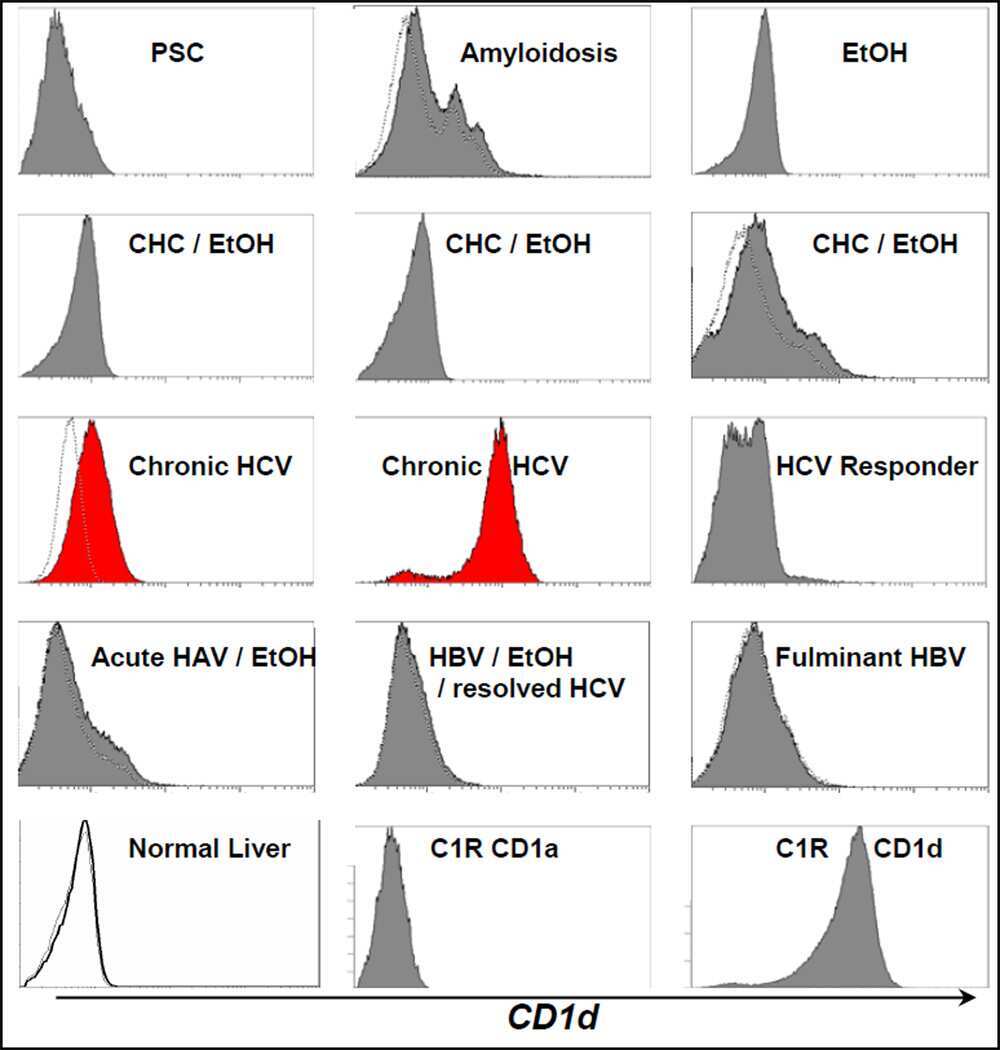
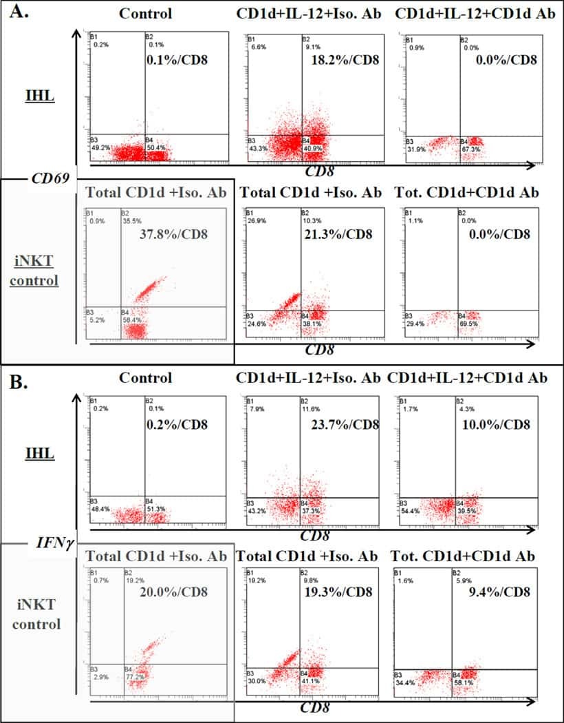
Ex vivo analysis of resident hepatic pro-inflammatory CD1d-reactive T cells and hepatocyte surface CD1d expression in hepatitis C.
Schistosomes induce regulatory features in human and mouse CD1d(hi) B cells: inhibition of allergic inflammation by IL-10 and regulatory T cells.
Joosten SA, van Meijgaarden KE, Del Nonno F, Baiocchini A, Petrone L, Vanini V, Smits HH, Palmieri F, Goletti D, Ottenhoff TH
PLoS pathogens 2016 Jun;12(6):e1005687
PLoS pathogens 2016 Jun;12(6):e1005687
Expression of CD1a and Type-1 Polarization Are Dissociated in Human Monocyte-Derived Dendritic Cells.
Mester B, Bauer E, Wood CE, Hermans IF, Gasser O
PloS one 2015;10(10):e0140432
PloS one 2015;10(10):e0140432
CD1d levels in peripheral blood of patients with acute myeloid leukemia and acute lymphoblastic leukemia.
Guo W, Dong A, Xing C, Lin X, Pan X, Lin Y, Zhu B, He M, Yao RX
Oncology letters 2014 Aug;8(2):825-830
Oncology letters 2014 Aug;8(2):825-830
Ex vivo analysis of resident hepatic pro-inflammatory CD1d-reactive T cells and hepatocyte surface CD1d expression in hepatitis C.
Yanagisawa K, Yue S, van der Vliet HJ, Wang R, Alatrakchi N, Golden-Mason L, Schuppan D, Koziel MJ, Rosen HR, Exley MA
Journal of viral hepatitis 2013 Aug;20(8):556-65
Journal of viral hepatitis 2013 Aug;20(8):556-65
Schistosomes induce regulatory features in human and mouse CD1d(hi) B cells: inhibition of allergic inflammation by IL-10 and regulatory T cells.
van der Vlugt LE, Labuda LA, Ozir-Fazalalikhan A, Lievers E, Gloudemans AK, Liu KY, Barr TA, Sparwasser T, Boon L, Ngoa UA, Feugap EN, Adegnika AA, Kremsner PG, Gray D, Yazdanbakhsh M, Smits HH
PloS one 2012;7(2):e30883
PloS one 2012;7(2):e30883
No comments: Submit comment
Supportive validation
- Submitted by
- Invitrogen Antibodies (provider)
- Main image
- Experimental details
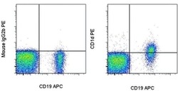
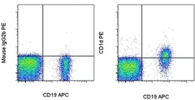
- Staining of normal human peripheral blood cells with Anti-Human CD19 APC (Product # 17-0199-42) and Mouse IgG2b K Isotype Control PE (Product # 12-4732-81) (left) or Anti-Human CD1d PE (right). Cells in the lymphocyte gate were used for analysis.
- Conjugate
- Yellow dye
Supportive validation
- Submitted by
- Invitrogen Antibodies (provider)
- Main image
- Experimental details
- NULL
- Conjugate
- Yellow dye
- Submitted by
- Invitrogen Antibodies (provider)
- Main image
- Experimental details
- NULL
- Conjugate
- Yellow dye