Explore
 Validate
 Learn
Learn Immunoprecipitation
 Flow cytometry
Antibody data
- Antibody Data
- Antigen structure
- References [9]
- Comments [0]
- Validations
- Flow cytometry [2]
- Other assay [8]
Submit
Validation data
Reference
Comment
Report error
- Product number
- 14-0016-37 - Provider product page

- Provider
- Invitrogen Antibodies
- Product name
- CD1d Monoclonal Antibody (51.1), eBioscience™
- Antibody type
- Monoclonal
- Antigen
- Other
- Description
- Description: The monoclonal antibody 51.1 reacts with human CD1d, a member of the CD1 family with similarity to the non-polymorphic MHC Class I-like molecules. CD1d is a highly conserved single transmembrane receptor of the Immunoglobulin Superfamily. CD1d can associate with beta-microglobulin another feature showing similarity to MHC class I molecules, but can also exist as a nonglycosylated protein not in association with beta microglobulin. This suggests different control mechanisms for presenting glycolipid containing molecules to CD1d reactive NKT cells. Expression of CD1d is found on B cells of the periphery, in resting monocytes and cortical thymocytes. On intestinal epithelial cells (IEC) expression is polarized. Expression can also be found at low levels intracellularly in hepatocytes. In HCV (hepatitis C virus) livers, CD1d is highly expressed compared to normal controls. The 51.1 monoclonal antibody has been shown to have functional activity; blocking the interaction of CD1d transfected cells with NKT cells. Applications Reported: This 51.1 antibody has been reported for use in flow cytometric analysis, immunoprecipitation, and immunohistology staining of frozen tissue sections. Applications Tested: This 51.1 antibody has been tested by flow cytometric analysis of normal human peripheral blood cells. This can be used at less than or equal to 0.5 µg per test. A test is defined as the amount (µg) of antibody that will stain a cell sample in a final volume of 100 µL. Cell number should be determined empirically but can range from 10^5 to 10^8 cells/test. It is recommended that the antibody be carefully titrated for optimal performance in the assay of interest. Purity: Greater than 90%, as determined by SDS-PAGE. Aggregation: Less than 10%, as determined by HPLC. Filtration: 0.2 µm post-manufacturing filtered.
- Reactivity
- Human
- Host
- Mouse
- Isotype
- IgG
- Antibody clone number
- 51.1
- Vial size
- 2 mg
- Concentration
- 0.5 mg/mL
- Storage
- 4°C
Submitted references Human CD40 ligand-expressing type 3 innate lymphoid cells induce IL-10-producing immature transitional regulatory B cells.
Leishmania infantum Exoproducts Inhibit Human Invariant NKT Cell Expansion and Activation.
Generation and characterization of CD1d-specific single-domain antibodies with distinct functional features.
Innate Invariant NKT Cell Recognition of HIV-1-Infected Dendritic Cells Is an Early Detection Mechanism Targeted by Viral Immune Evasion.
Potent neutralizing anti-CD1d antibody reduces lung cytokine release in primate asthma model.
Human cytomegalovirus (HCMV) US2 protein interacts with human CD1d (hCD1d) and down-regulates invariant NKT (iNKT) cell activity.
Ex vivo analysis of resident hepatic pro-inflammatory CD1d-reactive T cells and hepatocyte surface CD1d expression in hepatitis C.
Severe loss of invariant NKT cells exhibiting anti-HTLV-1 activity in patients with HTLV-1-associated disorders.
CD1d structure and regulation on human thymocytes, peripheral blood T cells, B cells and monocytes.
Komlósi ZI, Kovács N, van de Veen W, Kirsch AI, Fahrner HB, Wawrzyniak M, Rebane A, Stanic B, Palomares O, Rückert B, Menz G, Akdis M, Losonczy G, Akdis CA
The Journal of allergy and clinical immunology 2018 Jul;142(1):178-194.e11
The Journal of allergy and clinical immunology 2018 Jul;142(1):178-194.e11
Leishmania infantum Exoproducts Inhibit Human Invariant NKT Cell Expansion and Activation.
Belo R, Santarém N, Pereira C, Pérez-Cabezas B, Macedo F, Leite-de-Moraes M, Cordeiro-da-Silva A
Frontiers in immunology 2017;8:710
Frontiers in immunology 2017;8:710
Generation and characterization of CD1d-specific single-domain antibodies with distinct functional features.
Lameris R, de Bruin RC, van Bergen En Henegouwen PM, Verheul HM, Zweegman S, de Gruijl TD, van der Vliet HJ
Immunology 2016 Sep;149(1):111-21
Immunology 2016 Sep;149(1):111-21
Innate Invariant NKT Cell Recognition of HIV-1-Infected Dendritic Cells Is an Early Detection Mechanism Targeted by Viral Immune Evasion.
Paquin-Proulx D, Gibbs A, Bächle SM, Checa A, Introini A, Leeansyah E, Wheelock CE, Nixon DF, Broliden K, Tjernlund A, Moll M, Sandberg JK
Journal of immunology (Baltimore, Md. : 1950) 2016 Sep 1;197(5):1843-51
Journal of immunology (Baltimore, Md. : 1950) 2016 Sep 1;197(5):1843-51
Potent neutralizing anti-CD1d antibody reduces lung cytokine release in primate asthma model.
Nambiar J, Clarke AW, Shim D, Mabon D, Tian C, Windloch K, Buhmann C, Corazon B, Lindgren M, Pollard M, Domagala T, Poulton L, Doyle AG
mAbs 2015;7(3):638-50
mAbs 2015;7(3):638-50
Human cytomegalovirus (HCMV) US2 protein interacts with human CD1d (hCD1d) and down-regulates invariant NKT (iNKT) cell activity.
Han J, Rho SB, Lee JY, Bae J, Park SH, Lee SJ, Lee SY, Ahn C, Kim JY, Chun T
Molecules and cells 2013 Nov;36(5):455-64
Molecules and cells 2013 Nov;36(5):455-64
Ex vivo analysis of resident hepatic pro-inflammatory CD1d-reactive T cells and hepatocyte surface CD1d expression in hepatitis C.
Yanagisawa K, Yue S, van der Vliet HJ, Wang R, Alatrakchi N, Golden-Mason L, Schuppan D, Koziel MJ, Rosen HR, Exley MA
Journal of viral hepatitis 2013 Aug;20(8):556-65
Journal of viral hepatitis 2013 Aug;20(8):556-65
Severe loss of invariant NKT cells exhibiting anti-HTLV-1 activity in patients with HTLV-1-associated disorders.
Azakami K, Sato T, Araya N, Utsunomiya A, Kubota R, Suzuki K, Hasegawa D, Izumi T, Fujita H, Aratani S, Fujii R, Yagishita N, Kamijuku H, Kanekura T, Seino K, Nishioka K, Nakajima T, Yamano Y
Blood 2009 Oct 8;114(15):3208-15
Blood 2009 Oct 8;114(15):3208-15
CD1d structure and regulation on human thymocytes, peripheral blood T cells, B cells and monocytes.
Exley M, Garcia J, Wilson SB, Spada F, Gerdes D, Tahir SM, Patton KT, Blumberg RS, Porcelli S, Chott A, Balk SP
Immunology 2000 May;100(1):37-47
Immunology 2000 May;100(1):37-47
No comments: Submit comment
Supportive validation
- Submitted by
- Invitrogen Antibodies (provider)
- Main image

- Experimental details
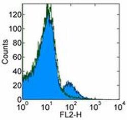
- Staining of normal human peripheral blood cells with 0.5 µg of Mouse IgG2b K Isotype Control Purified (Product # 14-4732-82) (open histogram) or Anti-Human CD1d Purified (filled histogram) followed by Anti-Mouse IgG Biotin (Product # 13-4013-85) and Streptavidin PE (Product # 12-4317-87). Cells in the lymphocyte gate were used for analysis.

- Submitted by
- Invitrogen Antibodies (provider)
- Main image
- Experimental details
- Staining of normal human peripheral blood cells with 0.5 µg of Mouse IgG2b K Isotype Control Purified (Product # 14-4732-82) (open histogram) or Anti-Human CD1d Purified (filled histogram) followed by Anti-Mouse IgG Biotin (Product # 13-4013-85) and Streptavidin PE (Product # 12-4317-87). Cells in the lymphocyte gate were used for analysis.

Supportive validation
- Submitted by
- Invitrogen Antibodies (provider)
- Main image

- Experimental details
- NULL
- Submitted by
- Invitrogen Antibodies (provider)
- Main image
- Experimental details
- NULL

- Submitted by
- Invitrogen Antibodies (provider)
- Main image
- Experimental details
- NULL

- Submitted by
- Invitrogen Antibodies (provider)
- Main image
- Experimental details
- NULL
- Submitted by
- Invitrogen Antibodies (provider)
- Main image

- Experimental details
- NULL
- Submitted by
- Invitrogen Antibodies (provider)
- Main image
- Experimental details
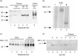
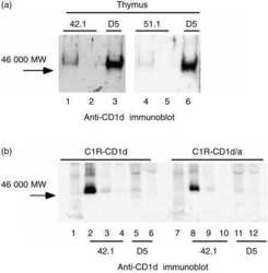
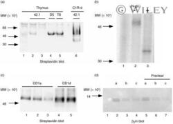
- 6 CD1d immunoprecipitations from thymocytes and specificity of D5 monoclonal antibody (mAb). (a) Streptavidin blot of immunoprecipitates from biotinylated thymocyte lysates: lane 1, normal mouse serum; lanes 2 and 3, sequential 42.1 anti-CD1d mAb precipitations; lane 4, D5 anti-CD1d mAb; lane 5, OKT6 anti-CD1a precipitation; lane 6, 42.1 precipitation from CD1d-transfected C1R cells. (b) Deglycosylation of CD1d from surface-iodinated thymocyte lysates: lane 1, normal mouse serum immunoprecipitation; lanes 2 and 3, D5 anti-CD1d mAb immunoprecipitates untreated (lane 2) or treated lane 3 with N -glycanase ( N -glc). (c) D5 immuno-precipitation from CD1a precleared biotinylated thymocyte lysate: lanes 1-4, lysate immunoprecipitated sequentially with OKT6 (anti-CD1a) beads (lanes 1-3) followed by reimmunoprecipitation with D5 (anti-CD1d) beads (lane 4); lane 5, D5 precipitation from an equal amount of lysate without a CD1a preclear. (d) Immunoprecipitates from unlabelled thymocyte lysates immunoblotted with BBM.1 anti-beta 2 m mAb: lanes 1-4, fresh thymocyte lysate; lanes 5-7, equivalent amounts of D5 anti-CD1d mAb-depleted thymocyte lysate. Immunoprecipitations were as follows: lane 1, normal mouse serum; lanes 2 and 5, anti-CD1a (OKT6) mAb; lanes 3 and 6, anti-CD1b (4A76) mAb; lanes 4 and 7, anti-CD1c (M241) mAb. Samples were analysed on a 15% gel under non-reducing conditions.

- Submitted by
- Invitrogen Antibodies (provider)
- Main image

- Experimental details
- Induction of a humoral immune response after immunization of two Lama glama with CD1d-transfected C1R cells. Reactivity of differentially diluted pre-immune (*), as well as post-immune day 28 (#) and day 43 (^) sera against whole C1R-CD1d cells as detected by flow cytometry after sequential incubation of C1R-CD1d cells with llama sera, rabbit-anti-llama sera and FITC-labelled swine anti-rabbit antibody. Data reflect mean fluorescence intensity (MFI) as determined in two individual llamas (a and b).

- Submitted by
- Invitrogen Antibodies (provider)
- Main image

- Experimental details
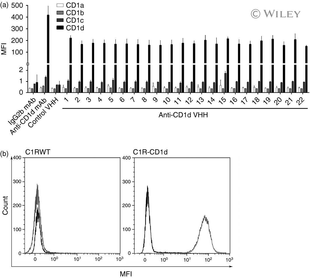
- Specificity of selected variable domain of heavy-chain-only antibodies (VHH) for CD1d and not CD1a, CD1b or CD1c. (a) Binding of the panel of purified anti-CD1d VHH clones, a non-specific negative control VHH, a positive control CD1d-specific monoclonal antibody (mAb), and a negative control IgG2b mAb against CD1a- (white bars), CD1b- (light grey bars), CD1c- (dark grey bars), and CD1d- (black bars) transfected cells. (b) Representative histograms showing binding of a negative control VHH (black) and anti-CD1d VHH21 (light grey) to wild-type C1R (C1R-WT, left histogram) and CD1d-transfected C1R cells (C1R-CD1d, right histogram). MFI was detected by flow cytometry after sequential incubation of CD1a-, CD1b-, CD1c-, and CD1d-transfected cells with purified VHH, anti-MYC mAb and allophycocyanin-labelled goat-anti-mouse Fab 2 fragment. Data represent mean + SD of three individual experiments.