Explore
 Validate
 Learn
Learn Western blot
Antibody data
- Antibody Data
- Antigen structure
- References [0]
- Comments [0]
- Validations
- Western blot [2]
- Immunocytochemistry [1]
- Immunohistochemistry [1]
- Flow cytometry [1]
Submit
Validation data
Reference
Comment
Report error
- Product number
- AP1494a - Provider product page

- Provider
- Abcepta
- Proper citation
- Abgent Cat#AP1494a, RRID:AB_1208520
- Product name
- CD19 Antibody (N-term)
- Antibody type
- Polyclonal
- Antigen
- Synthetic peptide
- Description
- Peptide Affinity Purified Rabbit Polyclonal Antibody (Pab)
- Reactivity
- Human
- Host
- Rabbit
- Isotype
- IgG
- Vial size
- 400 µl
- Concentration
- 0.5 mg/ml
- Storage
- Maintain refrigerated at 2-8°C for up to 6 months. For long term storage store at -20°C in small aliquots to prevent freeze-thaw cycles.
No comments: Submit comment
Supportive validation
- Submitted by
- Abcepta (provider)
- Main image
- Experimental details
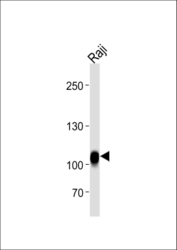
- CD19 Antibody (N-term) (Cat.# AP1494a) western blot analysis in Raji cell line lysates (35ug/lane).This demonstrates the CD19 antibody detected the CD19 protein (arrow).
- Primary Ab dilution
- 1:1000

- Submitted by
- Abcepta (provider)
- Main image

- Experimental details
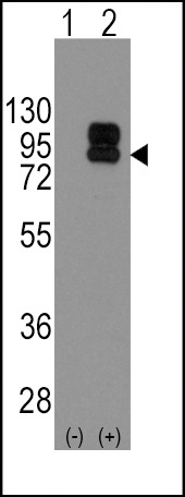
- Western blot analysis of CD19 (arrow) using rabbit polyclonal CD19 Antibody (N-term) (Cat.#AP1494a). 293 cell lysates (2 ug/lane) either nontransfected (Lane 1) or transiently transfected with the CD19 gene (Lane 2) (Origene Technologies).
- Primary Ab dilution
- 1:1000
Supportive validation
- Submitted by
- Abcepta (provider)
- Main image
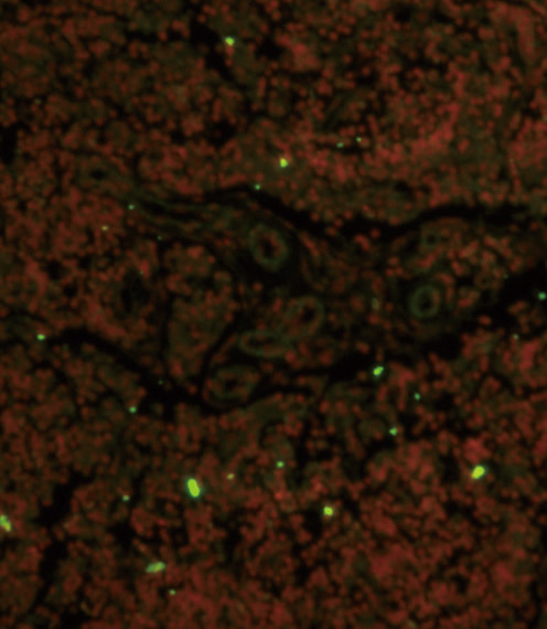
- Experimental details
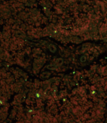
- Immunofluorescence analysis of CD19 Antibody (N-term) with paraffin-embedded human lymph tissue . 0.05 mg/ml primary antibody was followed by FITC-conjugated goat anti-rabbit lgG (whole molecule). FITC emits green fluorescence.Red counterstaining is PI.
- Primary Ab dilution
- 1:10~50
Supportive validation
- Submitted by
- Abcepta (provider)
- Main image

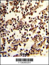
- Experimental details
- "Formalin-fixed and paraffin-embedded human lymph reacted with CD19 Antibody (N-term), which was peroxidase-conjugated to the secondary antibody, followed by DAB staining. This data demonstrates the use of this antibody for immunohistochemistry; clinical relevance has not been evaluated."
- Primary Ab dilution
- 1:10~50
Supportive validation
- Submitted by
- Abcepta (provider)
- Main image
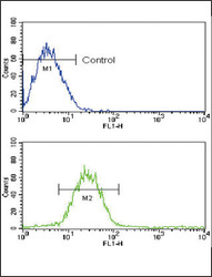
- Experimental details
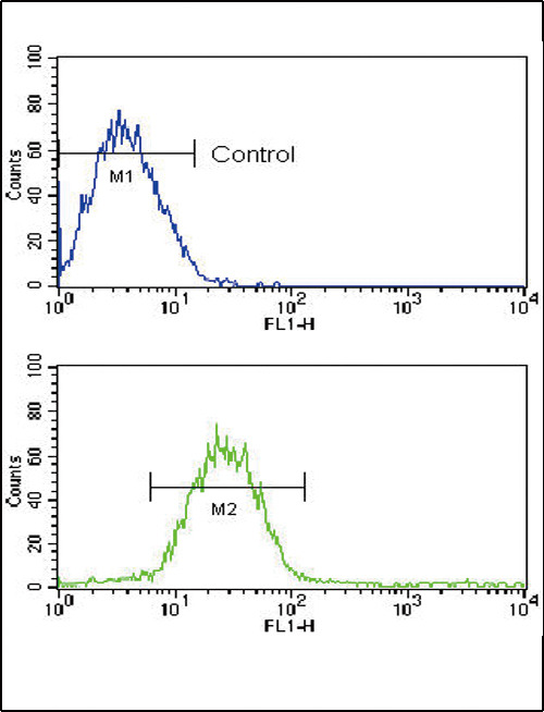
- Flow cytometric analysis of CEM cells using CD19 Antibody (N-term)(bottom histogram) compared to a negative control cell (top histogram). FITC-conjugated goat-anti-rabbit secondary antibodies were used for the analysis.
- Primary Ab dilution
- 1:10~50