 Explore
 Validate
 Learn
Learn Western blot
 ELISA
ELISAAntibody data
- Antibody Data
- Antigen structure
- References [0]
- Comments [0]
- Validations
- Western blot [3]
- Immunocytochemistry [1]
- Immunohistochemistry [1]
- Flow cytometry [1]
Submit
Validation data
Reference
Comment
Report error
- Product number
- F44318 - Provider product page

- Provider
- NSJ Bioreagents
- Product name
- CD19 Antibody
- Antibody type
- Polyclonal
- Description
- This highly specific CD19 antibody is suitable for use in Western blot/Immunofluorescence/Flow cytometry/Immunohistochemistry/ELISA applications with human samples.
- Reactivity
- Human
- Host
- Rabbit
- Conjugate
- Unconjugated
- Vial size
- 0.08 ml, 0.4 ml
- Concentration
- In 1X PBS, pH 7.4, with 0.09% sodium azide
- Storage
- Aliquot the CD19 antibody and store frozen at -20oC or colder. Avoid repeated freeze-thaw cycles.
No comments: Submit comment
Supportive validation
- Submitted by
- NSJ Bioreagents (provider)
- Main image
- Experimental details
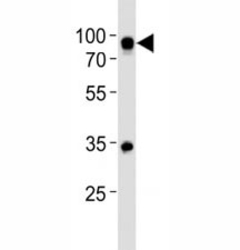
- Western blot analysis of lysate from Ramos cell line using CD19 antibody at 1:500. Expected size is 60~100 kDa depending on glycosylation level.

- Submitted by
- NSJ Bioreagents (provider)
- Main image
- Experimental details
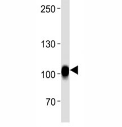
- CD19 antibody western blot analysis in Raji lysate. Expected size is 60~100 kDa depending on glycosylation level.

- Submitted by
- NSJ Bioreagents (provider)
- Main image
- Experimental details
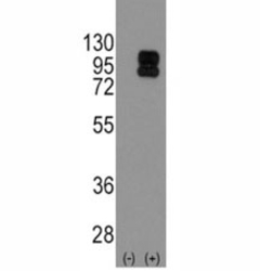
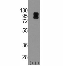
- Western blot analysis of CD19 antibody and 293 cell lysate (2 ug/lane) either nontransfected (Lane 1) or transiently transfected with the CD19 gene (2). Expected size is 60~100 kDa depending on glycosylation level.
Supportive validation
- Submitted by
- NSJ Bioreagents (provider)
- Main image
- Experimental details
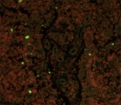
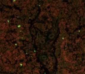
- Immunofluorescence analysis of CD19 antibody with paraffin-embedded human lymph tissue. Primary Ab was followed by FITC-conjugated goat anti-rabbit lgG (whole molecule). FITC emits green fluorescence. Red counterstaining is PI.
Supportive validation
- Submitted by
- NSJ Bioreagents (provider)
- Main image
- Experimental details
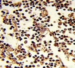
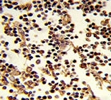
- IHC analysis of FFPE human lymph stained with CD19 antibody
Supportive validation
- Submitted by
- NSJ Bioreagents (provider)
- Main image
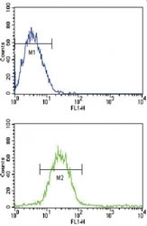
- Experimental details
- Flow cytometric analysis of CEM cells using CD19 antibody (bottom histogram) compared to a negative control cell (top histogram). FITC-conjugated goat-anti-rabbit secondary Ab was used for the analysis.
