Explore
 Validate
 Learn
Learn Western blot
 Immunocytochemistry
Antibody data
- Antibody Data
- Antigen structure
- References [7]
- Comments [0]
- Validations
- Immunocytochemistry [1]
- Immunohistochemistry [3]
- Other assay [5]
Submit
Validation data
Reference
Comment
Report error
- Product number
- 14-0194-37 - Provider product page

- Provider
- Invitrogen Antibodies
- Product name
- CD19 Monoclonal Antibody (6OMP31), eBioscience™
- Antibody type
- Monoclonal
- Antigen
- Other
- Description
- Description: The 6OMP31 monoclonal antibody reacts with human, mouse, and rat CD19. CD19 is a transmembrane glycoprotein belonging to the immunoglobulin superfamily. CD19 was first identified as the B4 antigen of human B lymphocytes using the anti-B4 monoclonal. It is expressed by B cells during all stages of development, except on terminally differentiated plasma cells, and on follicular dendritic cells. CD19 acts primarily as a B cell co-receptor and together with CD21, CD81, Leu13, and MHC class II forms a multi-molecular complex that associates with the BCR. CD19 plays a role in the development, activation, differentiation, and proliferation of B cells. CD19 deficiency leads to an overall impaired humoral immune response. Mutations in CD19 have been associated with severe immunodeficiency syndromes. Although it is unclear whether CD19 contributes directly to B cell carcinogenesis, it is expressed on most B cell tumors, such as acute lymphoblastic leukemias and B cell lymphomas. The use of CD19 monoclonal antibodies for therapies against lymphoma, leukemia and autoimmune disorders are currently being explored. Applications Reported: This 6OMP31 antibody has been reported for use in western blotting, immunohistochemical staining of frozen tissue sections, and immunohistochemical staining of formalin-fixed paraffin embedded tissue sections. Applications Tested: This 6OMP31 antibody has been tested by immunohistochemistry of formalin-fixed paraffin embedded tissue using low or high pH antigen retrieval and can be used at less than or equal to 0.5 µg/mL. The 6OMP31 antibody has been tested by immunohistochemstry of frozen tissue and can be used at less than or equal to 0.5 µg/mL. It is recommended that the antibody be carefully titrated for optimal performance in the assay of interest. Purity: Greater than 90%, as determined by SDS-PAGE. Aggregation: Less than 10%, as determined by HPLC. Filtration: 0.2 µm post-manufacturing filtered.
- Reactivity
- Human, Mouse, Rat
- Host
- Rat
- Isotype
- IgG
- Antibody clone number
- 6OMP31
- Vial size
- 2 mg
- Concentration
- 0.5 mg/mL
- Storage
- 4°C
Submitted references Knockdown of CD146 promotes endothelial-to-mesenchymal transition via Wnt/β-catenin pathway.
Fecal microbiota transplantation ameliorates atherosclerosis in mice with C1q/TNF-related protein 9 genetic deficiency.
Distribution of Immune Cells Including Macrophages in the Human Cochlea.
Innate-like self-reactive B cells infiltrate human renal allografts during transplant rejection.
Single-cell evaluation reveals shifts in the tumor-immune niches that shape and maintain aggressive lesions in the breast.
Kynurenine plays an immunosuppressive role in 2,4,6-trinitrobenzene sulfate-induced colitis in mice.
Microsomal Prostaglandin E Synthase-1 Facilitates an Intercellular Interaction between CD4⁺ T Cells through IL-1β Autocrine Function in Experimental Autoimmune Encephalomyelitis.
Zhang ZY, Zhai C, Yang XY, Li HB, Wu LL, Li L
PloS one 2022;17(8):e0273542
PloS one 2022;17(8):e0273542
Fecal microbiota transplantation ameliorates atherosclerosis in mice with C1q/TNF-related protein 9 genetic deficiency.
Kim ES, Yoon BH, Lee SM, Choi M, Kim EH, Lee BW, Kim SY, Pack CG, Sung YH, Baek IJ, Jung CH, Kim TB, Jeong JY, Ha CH
Experimental & molecular medicine 2022 Feb;54(2):103-114
Experimental & molecular medicine 2022 Feb;54(2):103-114
Distribution of Immune Cells Including Macrophages in the Human Cochlea.
Liu W, Danckwardt-Lillieström N, Schrott-Fischer A, Glueckert R, Rask-Andersen H
Frontiers in neurology 2021;12:781702
Frontiers in neurology 2021;12:781702
Innate-like self-reactive B cells infiltrate human renal allografts during transplant rejection.
Asano Y, Daccache J, Jain D, Ko K, Kinloch A, Veselits M, Wolfgeher D, Chang A, Josephson M, Cunningham P, Tambur A, Khan AA, Pillai S, Chong AS, Clark MR
Nature communications 2021 Jul 16;12(1):4372
Nature communications 2021 Jul 16;12(1):4372
Single-cell evaluation reveals shifts in the tumor-immune niches that shape and maintain aggressive lesions in the breast.
Sinha VC, Rinkenbaugh AL, Xu M, Zhou X, Zhang X, Jeter-Jones S, Shao J, Qi Y, Zebala JA, Maeda DY, McAllister F, Piwnica-Worms H
Nature communications 2021 Aug 18;12(1):5024
Nature communications 2021 Aug 18;12(1):5024
Kynurenine plays an immunosuppressive role in 2,4,6-trinitrobenzene sulfate-induced colitis in mice.
Tashita C, Hoshi M, Hirata A, Nakamoto K, Ando T, Hattori T, Yamamoto Y, Tezuka H, Tomita H, Hara A, Saito K
World journal of gastroenterology 2020 Mar 7;26(9):918-932
World journal of gastroenterology 2020 Mar 7;26(9):918-932
Microsomal Prostaglandin E Synthase-1 Facilitates an Intercellular Interaction between CD4⁺ T Cells through IL-1β Autocrine Function in Experimental Autoimmune Encephalomyelitis.
Takemiya T, Takeuchi C, Kawakami M
International journal of molecular sciences 2017 Dec 19;18(12)
International journal of molecular sciences 2017 Dec 19;18(12)
No comments: Submit comment
Supportive validation
- Submitted by
- Invitrogen Antibodies (provider)
- Main image
- Experimental details
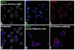
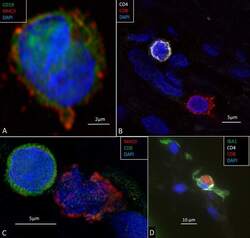
- Immunofluorescence analysis of B-lymphocyte antigen CD19 was performed using 70% confluent log phase Ramos cells. The cells were fixed with 4% paraformaldehyde for 10 minutes, permeabilized with 0.1% Triton™ X-100 for 15 minutes, and blocked with 2% BSA for 45 minutes at room temperature. The cells were labeled with CD19 Monoclonal Antibody (6OMP31), eBioscience™ (Product # 14-0194-82, 14-0194-80) at 5 µg/mL in 0.1% BSA, incubated at 4 degree celsius overnight and then labeled with Goat anti-Rat IgG (H+L) Cross-Adsorbed Secondary Antibody, Alexa Fluor 488 (Product # A11006), (1:2000 dilution), for 45 minutes at room temperature (Panel a: Green). Nuclei (Panel b: Blue) were stained with ProLong™ Diamond Antifade Mountant with DAPI (Product # P36962). F-actin (Panel c: Red) was stained with Rhodamine Phalloidin (Product # R415, 1:300 dilution). Panel d represents the merged image showing membrane localization. Panel e represents no expression of CD19 in Jurkat cells .Panel f represents control cells with no primary antibody to assess background. The images were captured at 60X magnification.

Supportive validation
- Submitted by
- Invitrogen Antibodies (provider)
- Main image
- Experimental details
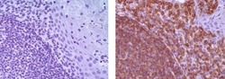
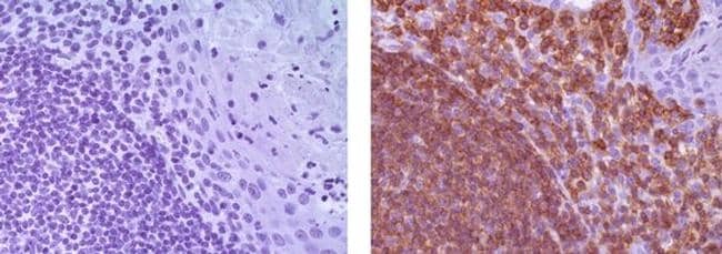
- Immunohistochemistry of formalin-fixed paraffin embedded human tonsil using 0.5 µg/mL Rat IgG2a K Isotype Control Purified (left) or 0.5 µg/mL Anti-CD19 Purified (right) followed by Anti-Rat IgG Biotin, Streptavidin HRP and DAB visualization. Nuclei are counterstained with hematoxylin.
- Submitted by
- Invitrogen Antibodies (provider)
- Main image
- Experimental details
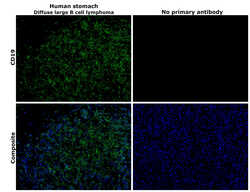
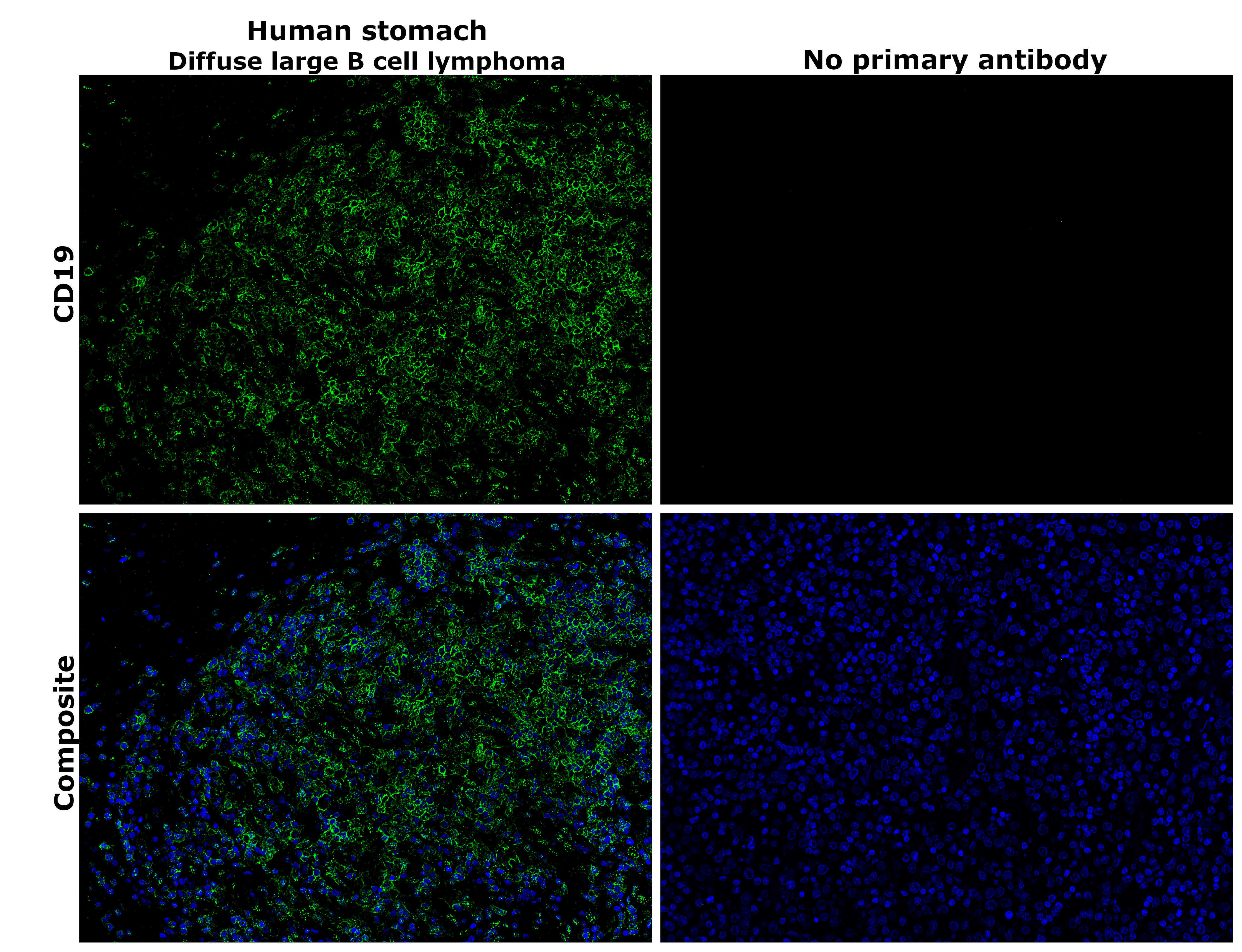
- Immunohistochemical analysis of CD19 was performed using formalin-fixed paraffin-embedded human stomach (Diffuse large B cell lymphoma) tissue sections. To expose the target protein, heat-induced epitope retrieval was performed on de-paraffinized sections using eBioscience™ IHC Antigen Retrieval Solution - Low pH (10X) (Product # 00-4955-58) diluted to 1X solution in water in a decloaking chamber at 110 degree Celsius for 15 minutes. Following antigen retrieval, the sections were blocked with 2% normal goat serum in 1X PBS for 45 minutes at room temperature and then probed with or without CD19 Monoclonal Antibody (6OMP31), eBioscience™ (Product # 14-0194-82) at 5 µg/mL in 0.1% normal goat serum overnight at 4 degree Celsius in a humidified chamber. Detection was performed using Goat anti-Rat IgG (H+L) Cross-Adsorbed Secondary Antibody, Alexa Fluor™ 488 (Product # A11006) at a dilution of 1:1,500 in 0.1% normal goat serum for 45 minutes at room temperature. ReadyProbes™ Tissue Autofluorescence Quenching Kit (Product # R37630) was used to quench autofluorescence from the tissues. Nuclei were stained with DAPI (Product # D1306) and the sections were mounted using ProLong™ Glass Antifade Mountant (Product # P36984). The images were captured on EVOS™ M7000 Imaging System (Product # AMF7000) at 20X magnification and externally deconvoluted.
- Submitted by
- Invitrogen Antibodies (provider)
- Main image
- Experimental details
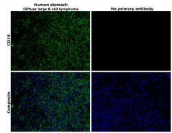
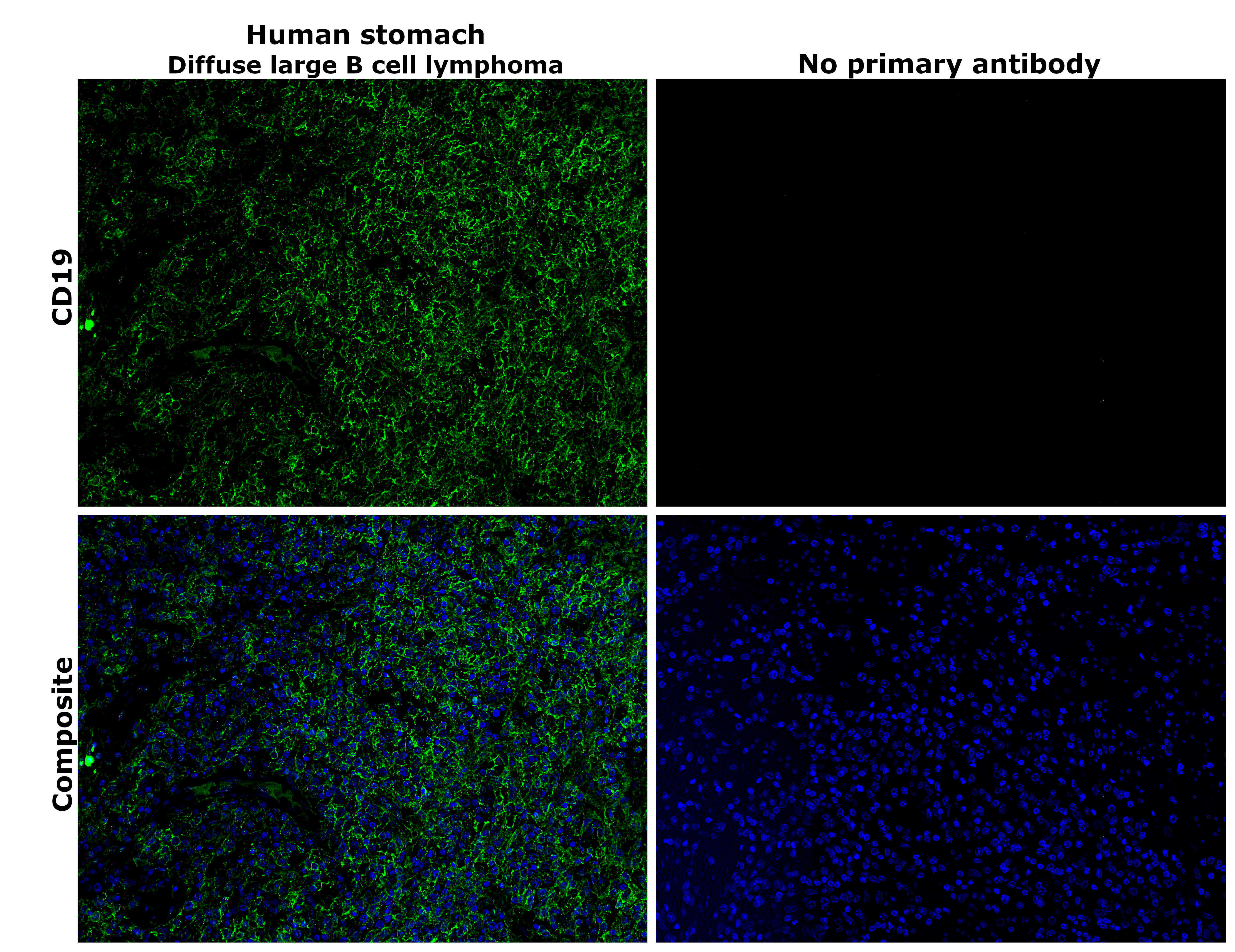
- Immunohistochemical analysis of CD19 was performed using formalin-fixed paraffin-embedded human stomach (Diffuse large B cell lymphoma) tissue sections. To expose the target protein, heat-induced epitope retrieval was performed on de-paraffinized sections using eBioscience™ IHC Antigen Retrieval Solution - High pH (10X) (Product # 00-4956-58) diluted to 1X solution in water in a decloaking chamber at 110 degree Celsius for 15 minutes. Following antigen retrieval, the sections were blocked with 2% normal goat serum in 1X PBS for 45 minutes at room temperature and then probed with or without CD19 Monoclonal Antibody (6OMP31), eBioscience™ (Product # 14-0194-82) at 5 µg/mL in 0.1% normal goat serum overnight at 4 degree Celsius in a humidified chamber. Detection was performed using Goat anti-Rat IgG (H+L) Cross-Adsorbed Secondary Antibody, Alexa Fluor™ 488 (Product # A11006) at a dilution of 1:1,500 in 0.1% normal goat serum for 45 minutes at room temperature. ReadyProbes™ Tissue Autofluorescence Quenching Kit (Product # R37630) was used to quench autofluorescence from the tissues. Nuclei were stained with DAPI (Product # D1306) and the sections were mounted using ProLong™ Glass Antifade Mountant (Product # P36984). The images were captured on EVOS™ M7000 Imaging System (Product # AMF7000) at 20X magnification and externally deconvoluted.
Supportive validation
- Submitted by
- Invitrogen Antibodies (provider)
- Main image
- Experimental details
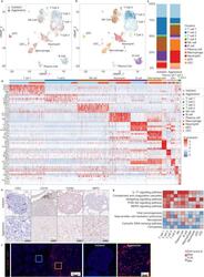
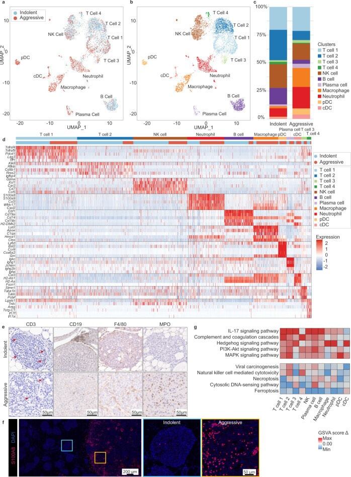
- Fig. 6 The aggressive niche harbors an expanded and spatially restricted myeloid population. a - c UMAP was used for dimension reduction of single-cell data. Immune fractions from indolent and aggressive lesions were computationally merged ( a ) for cluster analysis and identification ( b ). Proportion of clusters per group shown in c . NK natural killer, pDC plasmacytoid dendritic cell, cDC conventional dendritic cell. d Heatmap of top differentially expressed (DE) genes for each cluster, ordered in decreasing cluster size. Clusters indicated by top horizontal bar, with inferred immune identity noted. Cells derived from indolent or aggressive tumor sample indicated by second horizontal bar. DE genes are listed on the left. e Immunohistochemical staining for major lymphoid (T cells--CD3; B cells--CD19) and myeloid (macrophages--F4/80; neutrophils--MPO) populations associated with indolent and aggressive lesions. Arrows point to selected positively stained cells. Representative images shown from n = 4, 3, 3, 4 animals for CD3, CD19, F4/80, and MPO, respectively. f Immunofluorescent staining for neutrophils using an alternative marker S100A8. Left panel shows low power magnification view of adjacent indolent and aggressive lesions, with S100A8+ infiltration tightly localized with the aggressive niche. Boxes correspond by color to high magnification images shown middle (blue, indolent) and right (yellow, aggressive) panels. Representative images shown from n = 7 animals. g Immun
- Submitted by
- Invitrogen Antibodies (provider)
- Main image
- Experimental details
- Figure 13 (A) CD19-positive B lymphocyte in the modiolus. The cell also expresses MHCII. (B) CD4 and CD8-positive T cells in the peripheral region of the RC. (C) A CD8-positive lymphocyte and an MHCII-expressing cell in Rosenthal's canal. (D) A CD8 cell is seen to physically interact with an IBA1-positive macrophage.

- Submitted by
- Invitrogen Antibodies (provider)
- Main image
- Experimental details
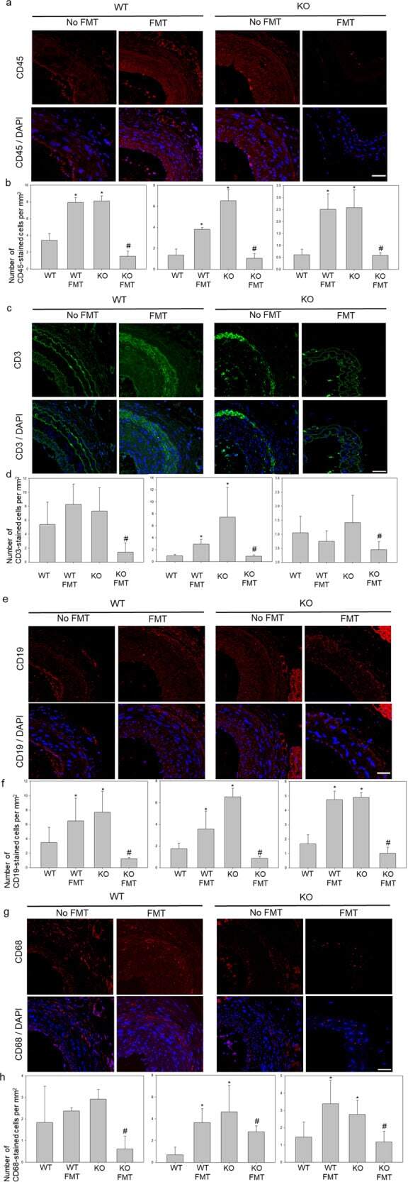
- The accumulation of immune cells results in atherosclerosis progression. a , b Immunofluorescence staining for the lymphocyte marker CD45 (red) and DAPI-stained nuclei (blue) are shown. c , d T cells were stained for CD3. e , f B cells were stained for CD19. g , h Macrophages were stained for CD68. * p < 0.05 for the WT-FMT group and the KO group compared to the WT group, # p < 0.05 for the KO-FMT group compared to the KO group. Two-way comparisons were performed with t tests. Scale bars in right lower corner represent 50 mum.
- Submitted by
- Invitrogen Antibodies (provider)
- Main image
- Experimental details
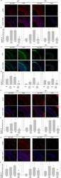
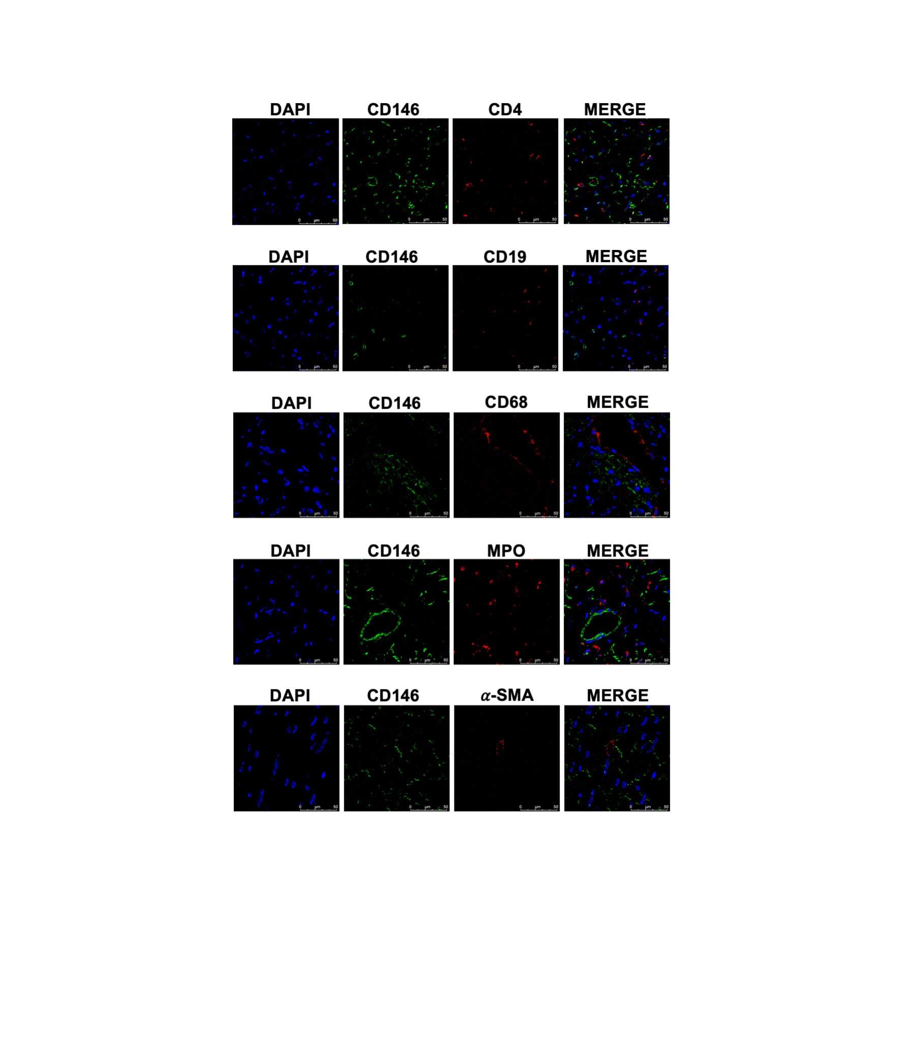
- S2 Fig CD146 was not expressed in SMCs and certain immune cells in adult mouse hearts. Representative double-immunofluorescence images of CD146 with alpha-SMA (a marker for SMCs), MPO (a marker for neutrophils), CD68 (a marker for monocytes/macrophages), CD19 (a marker for B lymphocytes), and CD4 (a marker for T-helper cells) in left ventricles from adult mice (scale bars indicate 50mum). Green represents CD146; red represents CD4, CD19, CD68, MPO and alpha-SMA, respectively; blue represents nuclei. (TIF) Click here for additional data file.
- Submitted by
- Invitrogen Antibodies (provider)
- Main image
- Experimental details
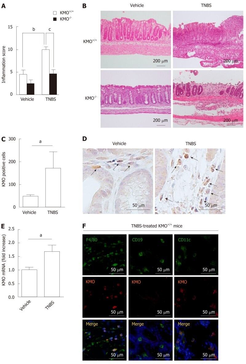
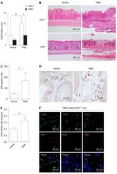
- Figure 1 Increased number of F4/80 + KMO + cells in the colon exacerbates TNBS-induced colitis. Mice were treated with either TNBS or vehicle, and colons were collected three days after treatment. A: Histological scores determined based on a histological grading as described in Materials and Methods. B: Representative images of H&E-stained colons of the said mice. C, D: The KMO + cells in the colons of the said mice were counted using immunohistochemical staining for KMO. E: KMO mRNA expression in the colon was determined by quantitative PCR. F: The colons of TNBS-treated mice were stained for F4/80, CD19, CD11c, KMO, and DAPI (nuclei) using immunofluorescence staining. The negative controls for F4/80, CD19, CD11c, and KMO were not stained ( Supplementary Figure 2 and 3 ). a P < 0.05 vs Vehicle group, b P < 0.01 vs Vehicle group, c P < 0.05 vs TNBS-treated KMO +/+ mice group. KMO: Kynurenine 3-monooxygenase; TNBS: Trinitrobenzene sulfonic acid.