Explore
 Validate
 Learn
Learn Western blot
 ELISA
ELISA Immunocytochemistry
Antibody data
- Antibody Data
- Antigen structure
- References [0]
- Comments [0]
- Validations
- Immunocytochemistry [3]
- Immunohistochemistry [1]
Submit
Validation data
Reference
Comment
Report error
- Product number
- MA5-15741 - Provider product page

- Provider
- Invitrogen Antibodies
- Product name
- CD19 Monoclonal Antibody (2E2B6B10)
- Antibody type
- Monoclonal
- Antigen
- Purifed from natural sources
- Description
- MA5-15741 targets CD19 in indirect ELISA, IHC and WB applications and shows reactivity with Human samples. The MA5-15741 immunogen is purified recombinant fragment of human CD19 expressed in E. Coli.
- Reactivity
- Human
- Host
- Mouse
- Isotype
- IgG
- Antibody clone number
- 2E2B6B10
- Vial size
- 100 μg
- Concentration
- Conc. Not Determined
- Storage
- Store at 4°C short term. For long term storage, store at -20°C, avoiding freeze/thaw cycles.
No comments: Submit comment
Supportive validation
- Submitted by
- Invitrogen Antibodies (provider)
- Main image

- Experimental details
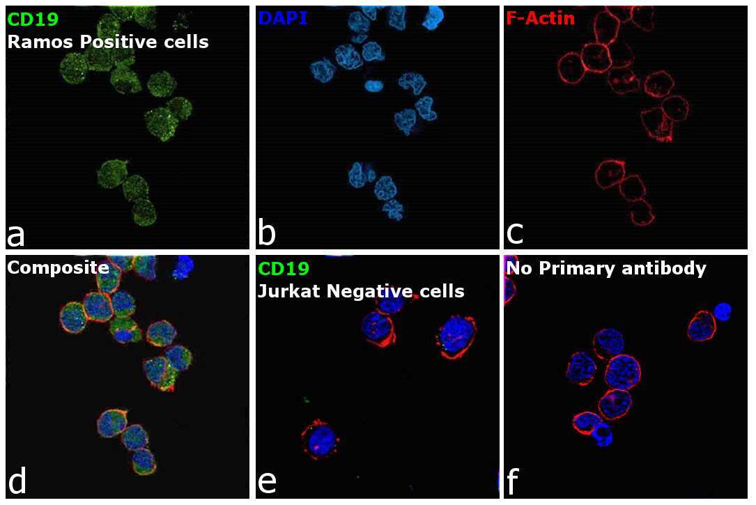
- Immunofluorescence analysis of B-lymphocyte antigen CD19 was performed using 70% confluent log phase Ramos cells. The cells were fixed with 4% paraformaldehyde for 10 minutes, permeabilized with 0.1% Triton™ X-100 for 15 minutes, and blocked with 2% BSA for 45 minutes at room temperature. The cells were labeled with CD19 Monoclonal Antibody (2E2B6B10) (Product # MA5-15741) at 1:100 dilution in 0.1% BSA, incubated at 4 degree celsius overnight and then labeled with Donkey anti-Mouse IgG (H+L) Highly Cross-Adsorbed Secondary Antibody, Alexa Fluor Plus 488 (Product # A32766), (1:2000 dilution), for 45 minutes at room temperature (Panel a: Green). Nuclei (Panel b:Blue) were stained with ProLong™ Diamond Antifade Mountant with DAPI (Product # P36962). F-actin (Panel c: Red) was stained with Rhodamine Phalloidin (Product # R415, 1:300 dilution). Panel d represents the merged image showing membrane as well as cytoplasmic localization. Panel e represents Jurkat cells having no expression of CD19. Panel f represents control cells with no primary antibody to assess background. The images were captured at 60X magnification. Immunofluorescence analysis of CD19 antibody was observed to be high in Ramos in comparison to low or negative expression in Jurkat . (doi :10.18632/oncotarget.24902).
- Submitted by
- Invitrogen Antibodies (provider)
- Main image

- Experimental details
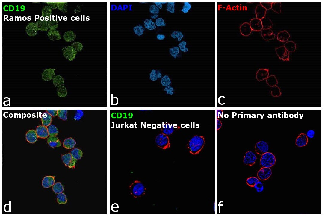
- Immunofluorescence analysis of B-lymphocyte antigen CD19 was performed using 70% confluent log phase Ramos cells. The cells were fixed with 4% paraformaldehyde for 10 minutes, permeabilized with 0.1% Triton™ X-100 for 15 minutes, and blocked with 2% BSA for 45 minutes at room temperature. The cells were labeled with CD19 Monoclonal Antibody (2E2B6B10) (Product # MA5-15741) at 1:100 dilution in 0.1% BSA, incubated at 4 degree celsius overnight and then labeled with Donkey anti-Mouse IgG (H+L) Highly Cross-Adsorbed Secondary Antibody, Alexa Fluor Plus 488 (Product # A32766), (1:2000 dilution), for 45 minutes at room temperature (Panel a: Green). Nuclei (Panel b:Blue) were stained with ProLong™ Diamond Antifade Mountant with DAPI (Product # P36962). F-actin (Panel c: Red) was stained with Rhodamine Phalloidin (Product # R415, 1:300 dilution). Panel d represents the merged image showing membrane as well as cytoplasmic localization. Panel e represents Jurkat cells having no expression of CD19. Panel f represents control cells with no primary antibody to assess background. The images were captured at 60X magnification. Immunofluorescence analysis of CD19 antibody was observed to be high in Ramos in comparison to low or negative expression in Jurkat . (doi :10.18632/oncotarget.24902).
- Submitted by
- Invitrogen Antibodies (provider)
- Main image

- Experimental details
- Immunofluorescence analysis of B-lymphocyte antigen CD19 was performed using 70% confluent log phase Ramos cells. The cells were fixed with 4% paraformaldehyde for 10 minutes, permeabilized with 0.1% Triton™ X-100 for 15 minutes, and blocked with 2% BSA for 45 minutes at room temperature. The cells were labeled with CD19 Monoclonal Antibody (2E2B6B10) (Product # MA5-15741) at 1:100 dilution in 0.1% BSA, incubated at 4 degree celsius overnight and then labeled with Donkey anti-Mouse IgG (H+L) Highly Cross-Adsorbed Secondary Antibody, Alexa Fluor Plus 488 (Product # A32766), (1:2000 dilution), for 45 minutes at room temperature (Panel a: Green). Nuclei (Panel b:Blue) were stained with ProLong™ Diamond Antifade Mountant with DAPI (Product # P36962). F-actin (Panel c: Red) was stained with Rhodamine Phalloidin (Product # R415, 1:300 dilution). Panel d represents the merged image showing membrane as well as cytoplasmic localization. Panel e represents Jurkat cells having no expression of CD19. Panel f represents control cells with no primary antibody to assess background. The images were captured at 60X magnification. Immunofluorescence analysis of CD19 antibody was observed to be high in Ramos in comparison to low or negative expression in Jurkat . (doi :10.18632/oncotarget.24902).

Supportive validation
- Submitted by
- Invitrogen Antibodies (provider)
- Main image

- Experimental details
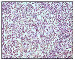
- Immunohistochemical analysis of paraffin-embedded human normal lymph node using CD19 monoclonal antibody (Product # MA5-15741) followed with DAB staining.