Explore
 Validate
 Learn
Learn Western blot
Antibody data
- Antibody Data
- Antigen structure
- References [0]
- Comments [0]
- Validations
- Western blot [1]
- Immunohistochemistry [1]
- Flow cytometry [1]
- Blocking/Neutralizing [1]
Submit
Validation data
Reference
Comment
Report error
- Product number
- AF3388 - Provider product page

- Provider
- R&D Systems
- Product name
- Human OX40/TNFRSF4 Antibody
- Antibody type
- Polyclonal
- Description
- Antigen Affinity-purified. Detects human OX40/TNFRSF4 in direct ELISAs and Western blots. In direct ELISAs, less than 5% cross-reactivity with recombinant mouse OX40 is observed.
- Reactivity
- Human
- Host
- Sheep
- Conjugate
- Unconjugated
- Antigen sequence
P43489- Isotype
- IgG
- Vial size
- 100 ug
- Concentration
- LYOPH
- Storage
- Use a manual defrost freezer and avoid repeated freeze-thaw cycles. 12 months from date of receipt, -20 to -70 °C as supplied. 1 month, 2 to 8 °C under sterile conditions after reconstitution. 6 months, -20 to -70 °C under sterile conditions after reconstitution.
No comments: Submit comment
Supportive validation
- Submitted by
- R&D Systems (provider)
- Main image

- Experimental details
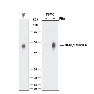
- Detection of Human OX40/TNFRSF4 by Western Blot. Western blot shows lysates of human T regulatory cells (Tregs) and human peripheral blood mononuclear cells (PBMCs) untreated (-) or treated (+) with 1 μg/mL PHA for 5 days. PVDF membrane was probed with 0.5 µg/mL of Sheep Anti-Human OX40/TNFRSF4 Antigen Affinity-purified Polyclonal Antibody (Catalog # AF3388) followed by HRP-conjugated Anti-Sheep IgG Secondary Antibody (Catalog # HAF016). A specific band was detected for OX40/TNFRSF4 at approximately 45 kDa (as indicated). This experiment was conducted under reducing conditions and using Immunoblot Buffer Group 1.
Supportive validation
- Submitted by
- R&D Systems (provider)
- Main image
- Experimental details
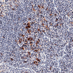
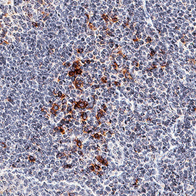
- OX40/TNFRSF4 in Human Tonsil. OX40/TNFRSF4 was detected in immersion fixed paraffin-embedded sections of human tonsil using Sheep Anti-Human OX40/TNFRSF4 Antigen Affinity-purified Polyclonal Antibody (Catalog # AF3388) at 3 µg/mL for 1 hour at room temperature followed by incubation with the Anti-Sheep IgG VisUCyte™ HRP Polymer Antibody (Catalog # VC006). Tissue was stained using DAB (brown) and counterstained with hematoxylin (blue). Specific staining was localized to plasma membrane. View our protocol for IHC Staining with VisUCyte HRP Polymer Detection Reagents.
Supportive validation
- Submitted by
- R&D Systems (provider)
- Main image
- Experimental details
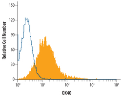
- Detection of OX40/TNFRSF4 in Human T Cells by Flow Cytometry. Human T cells were treated for 3-5 days with 1 μg/mL PHA then stained with Sheep Anti-Human OX40/ TNFRSF4 Antigen Affinity-purified Polyclonal Antibody (Catalog # AF3388, filled histogram) or control antibody (Catalog # 5-001-A, open histogram), followed by NorthernLights™ 557-conjugated Anti-Sheep IgG Secondary Antibody (Catalog # NL010).
Supportive validation
- Submitted by
- R&D Systems (provider)
- Main image
- Experimental details
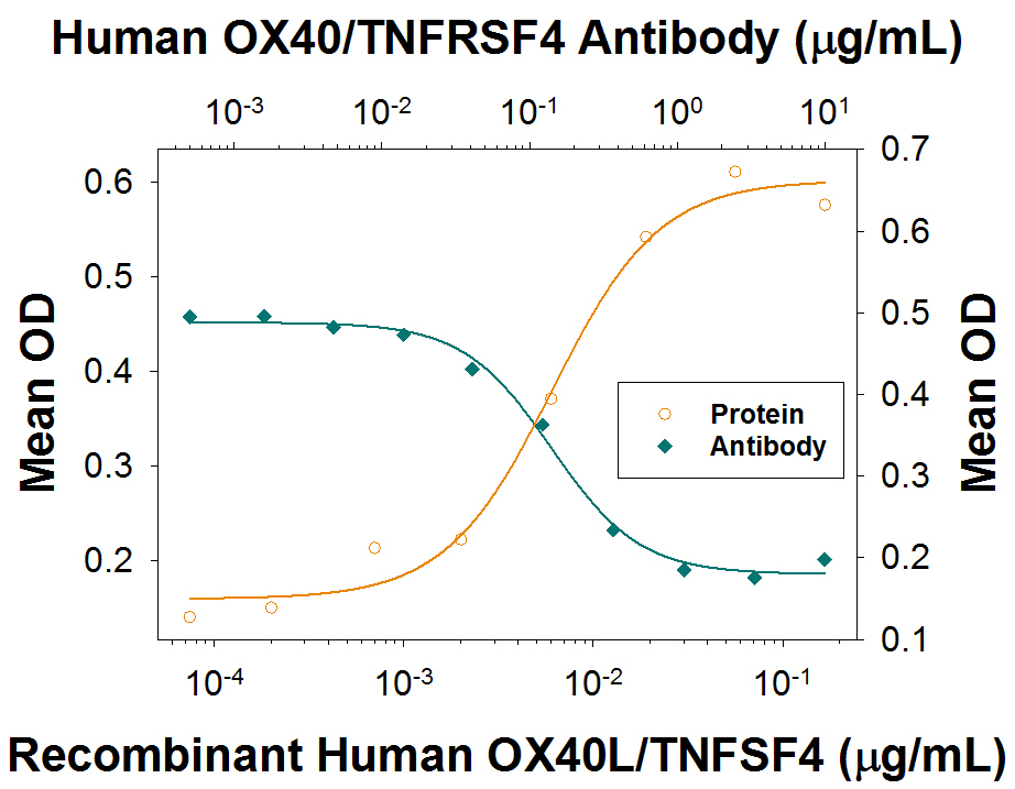
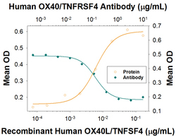
- CXCL8/IL-8 Secretion Induced by OX40L/TNFSF4 and Neutralization by Human OX40/TNFRSF4 Antibody. Recombinant Human OX40L/TNFSF4 (Catalog # 1054-OX) stimulates CXCL8/IL-8 secretion in the HT1080 human fibrosarcoma cell line transfected with human OX40/TNFRSF4 in a dose-dependent manner (orange line), as measured by the Human CXCL8/IL-8 Quantikine ELISA Kit (Catalog # D8000C). CXCL8/Il-8 Secretion elicited by Recombinant Human OX40L/TNFSF4 (10 ng/mL) is neutralized (green line) by increasing concentrations of Sheep Anti-Human OX40/TNFRSF4 Polyclonal Antibody (Catalog # AF3388). The ND50 is typically 0.025-0.16 µg/mL.