 Explore
 Validate
 Learn
Learn Western blot
Antibody data
- Antibody Data
- Antigen structure
- References [0]
- Comments [0]
- Validations
- Western blot [4]
- Immunocytochemistry [3]
Submit
Validation data
Reference
Comment
Report error
- Product number
- 701102 - Provider product page

- Provider
- Invitrogen Antibodies
- Product name
- GSK3B Recombinant Rabbit Monoclonal Antibody (1H9L3)
- Antibody type
- Monoclonal
- Antigen
- Recombinant full-length protein
- Description
- This antibody is predicted to react with non-human primate, mouse and rat based on sequence homology.
- Antibody clone number
- 1H9L3
- Concentration
- 0.5 mg/mL
No comments: Submit comment
Supportive validation
- Submitted by
- Invitrogen Antibodies (provider)
- Main image

- Experimental details
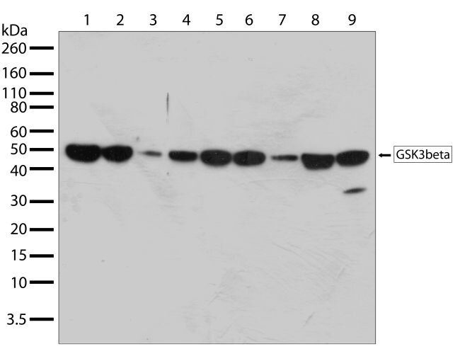
- Western blot analysis of GSK3 in whole cell extracts of HeLa (lane 1), HEK293 (lane 2), HepG2 (lane 3), Jurkat (lane 4), MCF-7 (lane 5), COLO 205 (lane 6), A431 (lane 7), COS7 (lane 8), and DU-145 (lane 9) using a GSK3 recombinant rabbit monoclonal antibody (Product # 701102) at a dilution of 0.1 µg/mL. Samples were detected using chemiluminescence (ECL). Results show a band at ~47kDa.
- Submitted by
- Invitrogen Antibodies (provider)
- Main image
- Experimental details
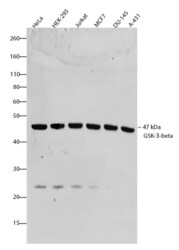
- Western blot analysis of GSK-3-beta was performed by loading 20 µg of HeLa, HEK-293, Jurkat, MCF-7, DU 145 and A431 cell lysates using Novex®NuPAGE®4-12% Bis-Tris gel (Product # NP0321BOX), XCell SureLock Electrophoresis System (Product # EI0002), Novex® Sharp Pre-Stained Protein Standard (Product # LC5800), and iBlot® Dry Blotting System (Product # IB21001). Proteins were transferred to a nitrocellulose membrane and blocked with 5% skim milk for 1 hour at room temperature. GSK-3-beta was detected at ~47 kDa using GSK-3-beta Recombinant Rabbit Monoclonal Antibody (Product # 701102) at a 1:1000 dilution in 2.5% skim milk at 4°C overnight on a rocking platform. Detection was performed using an HRP-conjugated Goat anti-Rabbit secondary antibody (Product # G-21234) at a 1:5000 dilution and chemiluminescent detection was performed using Pierce™ ECL Western blotting Substrate (Product # 32106).

- Submitted by
- Invitrogen Antibodies (provider)
- Main image
- Experimental details
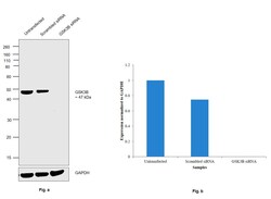
- Knockdown of Glycogen synthase kinase-3 beta was achieved by transfecting HeLa with Glycogen synthase kinase-3 beta specific siRNAs (Silencer® select Product # S6239, S6241). Western Blot analysis (Fig. a) was performed using Whole cell extracts from the Glycogen synthase kinase-3 beta knockdown cells (lane 3), non-targeting scrambled siRNA transfected cells (lane 2) and untransfected cells (lane 1). The Blot was probed with GSK3B Recombinant Rabbit Monoclonal Antibody (1H9L3) (Product # 701102, 1:1000 dilution ) and Goat anti-Rabbit IgG (H+L) Superclonal™ Recombinant Secondary Antibody, HRP (Product # A27036, 1:4000 dilution). Densitometric analysis of this western Blot is shown in histogram (Fig. b). Loss of signal upon siRNA mediated knock down confirms that antibody is specific to Glycogen synthase kinase-3 beta.

- Submitted by
- Invitrogen Antibodies (provider)
- Main image
- Experimental details
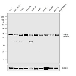
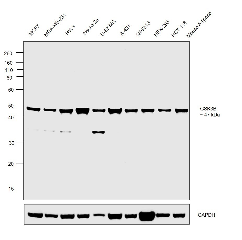
- Western Blot was performed using Anti-GSK3B Recombinant Rabbit Monoclonal Antibody (1H9L3) (Product # 701102) and a 47 kDa band corresponding to Glycogen synthase kinase-3 beta was observed across the cell lines and tissue tested. Whole cell extracts (30 µg lysate) of MCF7 (Lane 1), MDA-MB-231 (Lane 2), HeLa (Lane 3), Neuro-2a (Lane 4), U-87 MG (Lane 5), A-431 (Lane 6), NIH/3T3 (Lane 7), HEK-293 (Lane 8) and HCT 116 (Lane 9) and tissue extracts (30 µg lysate) of Mouse Adipose tissue (Lane 10) were electrophoresed using NuPAGE™ 10% Bis-Tris Protein Gel (Product # NP0302BOX). Resolved proteins were then transferred onto a Nitrocellulose membrane (Product # IB23001) by iBlot® 2 Dry Blotting System (Product # IB21001). The Blot was probed with the primary antibody (1:1000 dilution) and detected by chemiluminescence with Goat anti-Rabbit IgG (H+L) Superclonal™ Recombinant Secondary Antibody, HRP (Product # A27036, 1:4000 dilution) using the iBright FL 1000 (Product # A32752). Chemiluminescent detection was performed using Novex® ECL Chemiluminescent Substrate Reagent Kit (Product # WP20005).
Supportive validation
- Submitted by
- Invitrogen Antibodies (provider)
- Main image
- Experimental details
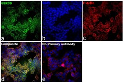
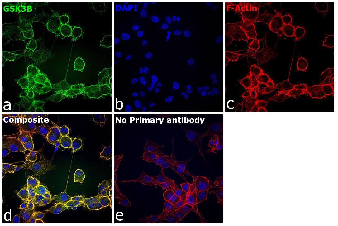
- Immunofluorescence analysis of GSK3B was performed using 70% confluent log phase U-2 OS cells. The cells were fixed with 4% paraformaldehyde for 10 minutes, permeabilized with 0.1% Triton™ X-100 for 15 minutes, and blocked with 2% BSA for 1 hour at room temperature. The cells were labeled with GSK3B Recombinant Rabbit Monoclonal Antibody (1H9L3) (Product # 701102) at 1:100 dilution in 0.1% BSA, incubated at 4°C overnight and then with Donkey anti-Rabbit IgG (H+L) Highly Cross-Adsorbed Secondary Antibody, Alexa Fluor Plus 488 (Product # A32790), (1:2,000 dilution) at a dilution of 1:2,000 for 45 minutes at room temperature (Panel a: green). Nuclei (Panel b: blue) were stained with Hoechst 33342 (Product # H1399). F-actin (Panel c: red) was stained with Rhodamine Phalloidin (Product # R415, 1:300). Panel d represents the merged image showing plasma membrane and cytoplasmic localization. Panel e represents control cells with no primary antibody to assess background. The images were captured at 40X magnification in CellInsight CX7 LZR High-Content Screening (HCS) Platform (Product # CX7C1115LZR).

- Submitted by
- Invitrogen Antibodies (provider)
- Main image

- Experimental details
- Immunofluorescence analysis of GSK3B was performed using 70% confluent log phase U-87 MG cells. The cells were fixed with 4% paraformaldehyde for 10 minutes, permeabilized with 0.1% Triton™ X-100 for 15 minutes, and blocked with 2% BSA for 1 hour at room temperature. The cells were labeled with GSK3B Recombinant Rabbit Monoclonal Antibody (1H9L3) (Product # 701102) at 1:100 dilution in 0.1% BSA, incubated at 4°C overnight and then with Donkey anti-Rabbit IgG (H+L) Highly Cross-Adsorbed Secondary Antibody, Alexa Fluor Plus 488 (Product # A32790), (1:2,000 dilution) at a dilution of 1:2,000 for 45 minutes at room temperature (Panel a: green). Nuclei (Panel b: blue) were stained with Hoechst 33342 (Product # H1399). F-actin (Panel c: red) was stained with Rhodamine Phalloidin (Product # R415, 1:300). Panel d represents the merged image showing plasma membrane and cytoplasmic localization. Panel e represents control cells with no primary antibody to assess background. The images were captured at 40X magnification in CellInsight CX7 LZR High-Content Screening (HCS) Platform (Product # CX7C1115LZR).
- Submitted by
- Invitrogen Antibodies (provider)
- Main image
- Experimental details
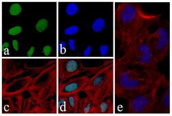
- Immunofluorescent analysis of GSK-3-beta was done on 70% confluent log phase U-2 OS cells. The cells were fixed with 4% paraformaldehyde for 15 minutes, permeabilized with 0.25% Triton X-100 for 10 minutes, and blocked with 5% BSA for 1 hour at room temperature. The cells were labeled with GSK-3-beta Recombinant Rabbit Monoclonal Antibody (Product # 701102) at a dilution of 1:400 in 1% BSA and incubated for 3 hours at room temperature and then labeled with Alexa Fluor® 488 Goat anti-Rabbit IgG Secondary Antibody (Product # A-11008) at a dilution of 1:400 for 30 minutes at room temperature (Panel a: green). Nuclei (Panel b: blue) were stained with SlowFade® Gold Antifade Mountant with DAPI (Product # S36938). F-actin (Panel c: red) was stained with Alexa Fluor® 594 Phalloidin (Product # A12381) and Panel d is a merged image showing nuclear localization. Panel e is a no primary antibody control. The images were captured using a Nikon microscope at 20X magnification.
